Imagine you are analyzing a urine sample for drug presence. Your instrument reads a specific concentration, but that number is wrong-not because the machine is broken, but because invisible components in the urine are blocking the signal. This phenomenon, known as ion suppression, which is a reduction in analyte signal caused by co-eluting matrix components in mass spectrometry, is one of the most persistent challenges in modern forensic and clinical toxicology. If left unchecked, it can lead to false negatives, underreported drug levels, and compromised legal or medical decisions.
In liquid chromatography-tandem mass spectrometry (LC-MS/MS), the gold standard for confirmatory testing, matrix effects occur when substances from biological samples like blood, plasma, or urine interfere with the ionization process. These interferences can suppress or enhance the signal of the target analyte, skewing results significantly. Understanding how to identify, measure, and mitigate these effects is not just a technical nicety; it is a regulatory requirement and a cornerstone of analytical integrity.
What Causes Ion Suppression?
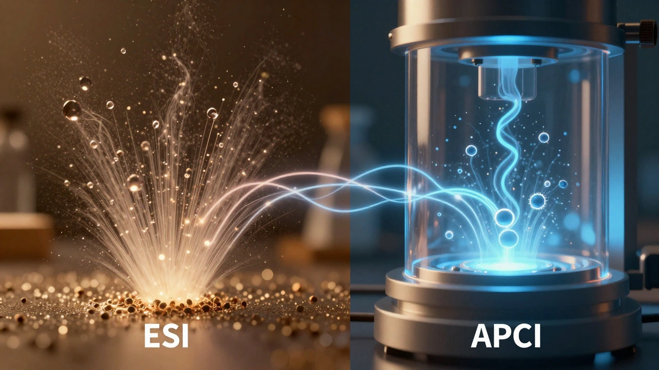
To grasp why ion suppression happens, you need to look at what occurs at the interface between the liquid chromatograph and the mass spectrometer. In electrospray ionization (ESI), the most common soft ionization technique used in toxicology, the sample solution is sprayed into tiny charged droplets. As these droplets evaporate, the analytes must reach the surface to be released as gas-phase ions into the vacuum of the mass spectrometer.
The problem arises when other compounds in your sample-collectively called the "matrix"-compete for this space. Biological matrices are messy. They contain proteins, phospholipids, salts (like sodium and chloride), urea, and endogenous metabolites. When these co-elute with your drug of interest, they can:
- Compete for charge at the droplet surface, pushing your analyte out.
- Bind to the analyte in non-ionizable complexes.
- Neutralize charged analytes before they can escape the droplet.
- Alter the physical properties of the droplet, hindering desolvation.
For instance, phospholipids in plasma are notorious for creating broad regions of suppression. They are surface-active molecules that accumulate in the ESI droplets, effectively shielding other compounds from ionization. Similarly, polyethylene glycol (PEG) oligomers found in urine can cause severe suppression for drugs that elute at similar retention times. A 2024 study on urine markers showed that drugs like MDMA, cocaine, and LSD experienced up to 90% signal loss when co-eluting with PEG 6-11 chains.
Measuring Matrix Effects: The Matuszewski Method
You cannot fix what you do not measure. Since the early 2000s, the framework proposed by Bogusław M. Matuszewski has been the industry standard for quantifying matrix effects. This method involves three distinct sets of samples:
- Neat Standard: The analyte dissolved in pure solvent (no matrix).
- Post-Extraction Spike: Blank matrix extract spiked with the analyte after extraction.
- Pre-Extraction Spike: Matrix spiked with the analyte before extraction.
By comparing the peak areas of these samples, you calculate two critical metrics:
- Matrix Effect (ME): Calculated as 100 × (Post-Extraction / Neat) − 100. A value of 0% means no effect. Negative values indicate suppression; positive values indicate enhancement.
- Extraction Recovery (RE): Calculated as 100 × (Pre-Extraction / Post-Extraction). This tells you how efficiently your sample prep method pulls the drug out of the matrix.
Regulatory guidelines from the U.S. FDA and European Medicines Agency require evaluating matrix effects using at least six individual lots of blank matrix to account for biological variability. If your ME varies wildly between samples, your method is not robust enough for routine use.

Strategies to Overcome Ion Suppression
Mitigating matrix effects requires a layered approach. No single trick works for every scenario, so you must combine sample preparation, chromatographic optimization, and intelligent calibration.
1. Sample Preparation Optimization
The goal here is to remove interferents while retaining the analyte. Common techniques include:
- Solid-Phase Extraction (SPE): Mixed-mode or polymeric sorbents can selectively retain drugs while washing away salts and proteins. Specialized phospholipid-removal cartridges are particularly effective for plasma samples.
- Liquid-Liquid Extraction (LLE): Using immiscible organic solvents to partition analytes away from aqueous matrix components. However, LLE may not remove all amphiphilic interferents like certain polymers.
- Dilution: Sometimes, less is more. Diluting the sample 1:10 or 1:20 with mobile phase can reduce the concentration of suppressing species below their threshold of interference. This "dilute-and-shoot" approach is gaining popularity for high-concentration targets where sensitivity is not a limiting factor.
2. Chromatographic Separation
If you cannot remove the interferent, separate it from the analyte. Adjusting your LC gradient, changing column chemistry (e.g., switching from C18 to phenyl-hexyl), or extending run times can move major endogenous peaks away from your target drugs. For example, shifting the elution window of polar compounds away from the "void volume" where salts and lipids often cluster can dramatically improve signal stability.
3. Ionization Mode Selection
Not all ionization sources are equal regarding matrix susceptibility. Electrospray Ionization (ESI) is highly sensitive to matrix effects because it relies on droplet surface phenomena. Atmospheric Pressure Chemical Ionization (APCI), on the other hand, uses gas-phase ion-molecule reactions and is generally more robust against phospholipid suppression. For non-polar, thermally stable analytes like steroid hormones, switching from ESI to APCI can eliminate many matrix-related issues.
4. Internal Standards and Calibration
This is your last line of defense. Using a stable isotope-labeled internal standard (SIL-IS) is crucial. The SIL-IS should be structurally identical to the analyte but heavier (e.g., labeled with Carbon-13 or Nitrogen-15 instead of Deuterium). Because it behaves identically during chromatography and ionization, it experiences the same suppression as the analyte. Quantification is then based on the ratio of analyte to internal standard, canceling out the matrix effect.
Critical Warning: Not all isotopic labels are created equal. Deuterium-labeled standards can sometimes exhibit slightly different retention times due to the "deuterium effect," leading to mismatched suppression profiles. Carbon-13 or Nitrogen-15 labels are preferred because they mimic the analyte's behavior more accurately. Always verify co-elution via post-column infusion experiments.
| Strategy | Pros | Cons | Best For |
|---|---|---|---|
| SPE Cleanup | Removes phospholipids/proteins | Time-consuming, higher cost | Plasma/Serum samples |
| Dilution | Fast, simple, low cost | Reduces sensitivity | High-concentration analytes |
| Chromatographic Shift | Separates interferents | May increase run time | Complex multi-analyte panels |
| APCI Source | Robust against lipids | Requires volatile/non-polar analytes | Steroids, lipophilic drugs |
| SIL-IS Calibration | Corrects residual effects | Expensive standards | All quantitative methods |
Real-World Implications in Forensic Toxicology
In a forensic setting, accuracy is paramount. A 50-90% suppression error can turn a positive case into a negative one, potentially releasing an impaired driver or exonerating someone who committed a crime under the influence. Conversely, ion enhancement can falsely inflate concentrations, leading to incorrect legal penalties.
Recent studies highlight the severity of this issue. In urinary biomarker analysis for solvent exposure, researchers observed ~50% suppression for methylhippuric acid derivatives due to co-eluting urine matrix. In another study involving drugs like tramadol and LSD, suppression reached ~90% when PEG oligomers were present. These are not edge cases; they are common occurrences in routine screening.
Laboratories must therefore adopt a proactive stance. Regular maintenance of the MS source, frequent evaluation of matrix effects across new batches of reagents, and continuous monitoring of internal standard responses are essential quality control measures. Software tools that flag abnormal IS response ratios can serve as an early warning system for matrix-induced bias.
Future Directions
As technology evolves, new solutions are emerging. Microflow and nanoflow LC systems produce smaller droplets, improving ionization efficiency and reducing the relative impact of matrix components. High-resolution accurate mass (HRAM) instruments offer better selectivity, allowing analysts to isolate analyte signals from chemical noise more effectively. Additionally, advanced software algorithms are beginning to normalize signals in real-time using endogenous reference ions, providing dynamic correction for matrix effects.
However, the fundamental principles remain unchanged. You must understand your matrix, prepare your samples intelligently, separate your analytes carefully, and calibrate rigorously. Ion suppression is not a bug to be patched; it is a characteristic of complex biological analysis that demands respect and systematic management.
What is the difference between ion suppression and ion enhancement?
Ion suppression occurs when matrix components reduce the ionization efficiency of the analyte, leading to a lower signal than expected. Ion enhancement is the opposite, where matrix components increase the ionization efficiency, resulting in a falsely high signal. Both are forms of matrix effects and can compromise quantitative accuracy.
Why are phospholipids such a big problem in LC-MS/MS?
Phospholipids are abundant in plasma and serum. They are surface-active molecules that accumulate in the electrospray droplets, competing with analytes for charge and space at the droplet surface. This leads to broad regions of ion suppression, particularly affecting moderately polar drugs.
How do I know if my internal standard is correcting for matrix effects?
You must verify that the internal standard co-elutes perfectly with the analyte and experiences the same degree of suppression. This is typically done using post-column infusion experiments. If the IS elutes even slightly earlier or later, it may not experience the same matrix environment, leading to biased quantitation.
Is dilution always a viable solution for matrix effects?
Dilution can be effective if the analyte concentration is high enough that the reduced sensitivity does not impact detection limits. However, for trace-level analysis, dilution may push the signal below the limit of quantitation (LOQ), making it unsuitable for low-abundance targets.
Which ionization source is less susceptible to matrix effects?
Atmospheric Pressure Chemical Ionization (APCI) is generally less susceptible to matrix effects than Electrospray Ionization (ESI), particularly for non-polar and thermally stable compounds. APCI relies on gas-phase reactions rather than droplet surface phenomena, making it more robust against phospholipid interference.