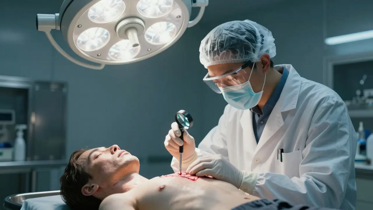
When a body arrives at the morgue, the clock is ticking. The pathologist has to figure out not just what killed the person, but how they died and when. This isn't about guessing; it's about reading the physical evidence left behind in tissue, bone, and blood. Wound analysis is the core skill that turns a silent body into a witness.
Every wound tells a story. A cut might look like an accident, but the angle could reveal a struggle. A bruise might seem minor, but its shape can identify the specific tool used. By understanding the mechanics of injury, forensic pathologists can distinguish between natural death, accident, suicide, and homicide. Here’s how they decode those stories.
The Three Main Categories of Trauma
Before diving into specifics, you need to know the big buckets. Forensic pathologists classify injuries primarily by the type of energy transferred to the body. There are three main categories: blunt force trauma, sharp force trauma, and firearm wounds. Each leaves distinct signatures that don’t lie.
- Blunt Force: Caused by objects with broad surfaces (fists, hammers, cars). These injuries crush or tear tissue rather than cutting it. Think bruises, lacerations, and fractures.
- Sharp Force: Caused by objects with edges (knives, glass, boxes). These injuries slice through tissue cleanly. Think incisions and punctures.
- Firearms: Caused by high-velocity projectiles. These involve complex interactions of entry, exit, and internal cavitation.
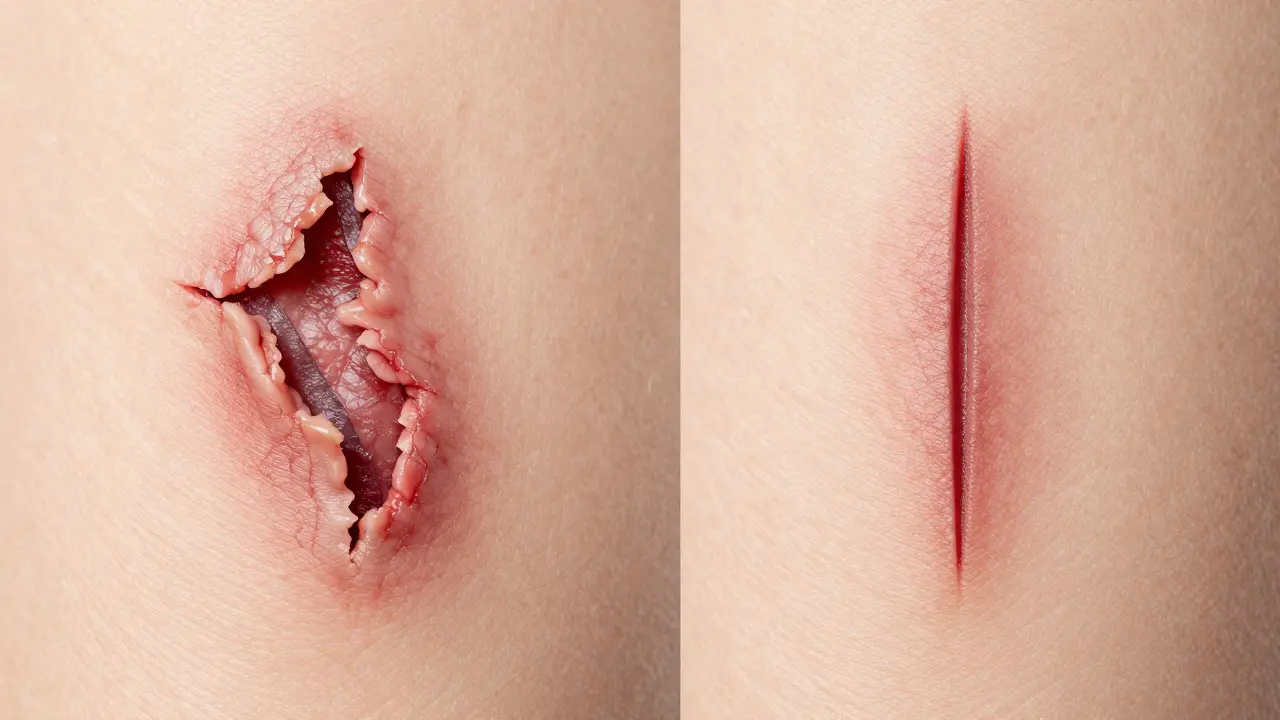
Misclassifying a wound is a common error in early investigations. For example, a deep laceration from a fall on concrete looks messy and jagged. An incision from a knife looks clean and straight. Confusing the two can lead investigators down the wrong path for days.
Reading Blunt Force Injuries
Blunt force is the most common cause of traumatic death outside of vehicle accidents. It’s also the trickiest because the skin tears, creating irregular edges that can mimic cuts. The key difference? Tension lines.
When blunt force hits the body, the underlying muscle and fat push against the skin, causing it to stretch and tear. This creates flaps of tissue inside the wound. If you see these flaps, it’s likely blunt force. If the edges are smooth and the depth matches the width consistently, it’s likely a sharp object.
Bruises are another major clue. A pathologist looks at the color, size, and shape of a bruise to estimate time since injury and the nature of the impact. A round, dark purple bruise suggests a direct hit from a fist. A linear, yellowish bruise might indicate a stick or rod. Even the pattern matters. If a victim falls onto a bedpost, the bruise will be vertical. If they were struck while standing, it might be horizontal. Context is everything.
| Feature | Blunt Force | Sharp Force |
|---|---|---|
| Wedge Edges | Jagged, irregular, torn tissue | Clean, smooth, continuous |
| Tissue Flaps | Present (internal tearing) | Absent (clean separation) |
| Depth Consistency | Varies widely | Uniform along length |
| Common Causes | Fists, hammers, vehicles | Knives, box cutters, glass |
Analyzing Sharp Force Wounds
Sharp force injuries are often more straightforward to analyze if the weapon is recovered. But what if the knife is missing? You have to reconstruct the blade from the wound itself.
Pathologists measure the maximum width of the wound. This gives them the minimum width of the blade. They also look at the length of the incision relative to the depth. A long, shallow cut suggests a slashing motion. A short, deep puncture suggests a stabbing motion. The angle of the entry point can even tell you where the attacker was standing relative to the victim.
One critical detail is the presence of defensive wounds. If a victim has multiple small cuts on their palms or forearms, it strongly suggests they tried to grab the weapon. This changes the narrative from a surprise attack to a struggle, which impacts legal proceedings significantly.
Decoding Firearm Wounds
Gunshot wounds are complex because they involve both mechanical damage and thermal effects. The pathologist must distinguish between entrance and exit wounds, as well as contact shots.
An entrance wound is typically smaller than the bullet diameter because the skin retracts after the bullet passes through. An exit wound is larger because the bullet expands and pushes tissue outward. Contact shots are unique. If the gun barrel touches the skin, gas and powder get injected under the skin, creating a star-shaped defect. This indicates the shot was fired at close range or point-blank.
Internal cavitation is another key factor. High-velocity bullets create a temporary cavity in the tissue, stretching organs and causing massive internal damage far beyond the path of the bullet. This explains why a small entrance hole can result in fatal organ rupture.
Determining Time Since Injury
Knowing when the injury happened is just as important as knowing how. Pathologists use biological markers to estimate the timeline. Bruises change color over time: red/purple immediately, then blue/green, then yellow/brown as hemoglobin breaks down. This progression takes about 10-14 days. However, this is only reliable if the body is fresh. Decomposition alters these colors rapidly.
For deeper tissues, pathologists look at histological changes. Inflammation starts within hours. Neutrophils arrive first, followed by macrophages. By examining the cellular response under a microscope, a pathologist can narrow down the time window to within a few days, even in decomposed bodies.
Common Pitfalls and Misinterpretations
Even experienced pathologists make mistakes. One common error is assuming all wounds are antemortem (before death). Postmortem changes can mimic injuries. Rigor mortis can pull muscles, creating false lacerations. Autolysis (self-digestion) can break down tissue, making old wounds look fresh. Always check for vital reactions-bleeding, inflammation, or healing signs-to confirm the injury happened while the person was alive.
Another pitfall is ignoring the context. A wound on the back of the head might look like a fall, but if there’s no corresponding fracture on the front, it could be a strike. Correlating external injuries with internal findings is crucial. A single data point is rarely enough.
FAQs About Wound Analysis
Can a pathologist tell exactly what weapon caused a wound?
Not always. They can determine the type of weapon (e.g., knife vs. axe) and sometimes the specific dimensions (e.g., blade width). Exact identification requires matching the wound to a recovered weapon. Without the weapon, they provide a profile of the likely instrument.
What is the difference between a laceration and an incision?
A laceration is a tear caused by blunt force, with ragged edges and tissue flaps. An incision is a cut caused by a sharp edge, with clean, smooth edges. The distinction is critical for determining the mechanism of injury.
How accurate is estimating time of death from wounds?
It varies. Early stages (first 24-48 hours) are more accurate due to clear biological changes. Later stages become less precise due to decomposition and environmental factors. Pathologists usually give a range, not a single hour.
Do all gunshot wounds have exit holes?
No. Low-velocity bullets or shots into dense bone may lodge the projectile, leaving no exit wound. The pathologist must examine the body thoroughly to find the bullet or track its path.
What role does microscopy play in wound analysis?
Microscopy allows pathologists to examine tissue structure at a cellular level. It helps identify subtle signs of vital reaction, such as hemorrhage around vessels, which confirms the injury occurred before death. It also reveals details invisible to the naked eye, like hair follicle damage or fiber imprints.