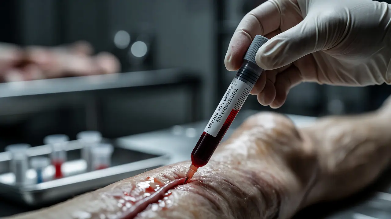
You’ve just finished the external examination. The body is on the table. You’re about to open it up. But before you make that first incision, there’s a critical question hanging in the air: how are you going to handle toxicology?
It sounds simple. Grab some blood, maybe some urine, send it off. But anyone who has worked in forensic pathology knows that toxicology sampling during an autopsy is where cases live or die. One contaminated vial, one wrong choice of preservative, or one missed window for gastric contents can turn a clear overdose case into an inconclusive mess. This isn’t just about following a checklist; it’s about understanding how drugs behave after death and making smart choices in real-time.
Why Standard Blood Draws Often Fail
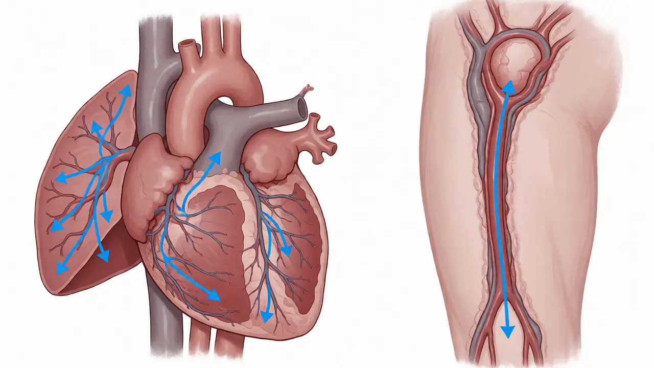
Here is the hard truth: peripheral blood is not always your best friend. When someone dies, circulation stops. Gravity takes over. Drugs redistribute from organs back into the blood vessels. This process, known as postmortem redistribution, can cause central blood levels (from the heart) to skyrocket compared to peripheral blood (from the femoral vein).
If you only take blood from the heart, you might think the person had a massive dose of fentanyl when they actually died of a natural cause with trace amounts in their system. If you only take peripheral blood, you might miss a lethal concentration if the sample was hemolyzed or clotted. The gold standard? Collect both. Always aim for femoral blood because it is less susceptible to contamination from the lungs and liver. It gives you a baseline that correlates better with antemortem levels.
The Essential Specimen Checklist
Don’t guess what the lab needs. Have a plan before you cut. While requirements vary by jurisdiction and specific drug classes, these are the non-negotiables for a comprehensive forensic toxicology screen.
- Femoral Blood: At least 10-20 mL. Use sodium fluoride/potassium oxalate tubes (gray top) to prevent glycolysis and preserve alcohol levels. Avoid heparin if possible, as it can interfere with some assays.
- Urine: If available, this is often more sensitive than blood for detecting recent use, especially for opioids and benzodiazepines. Even small amounts are valuable.
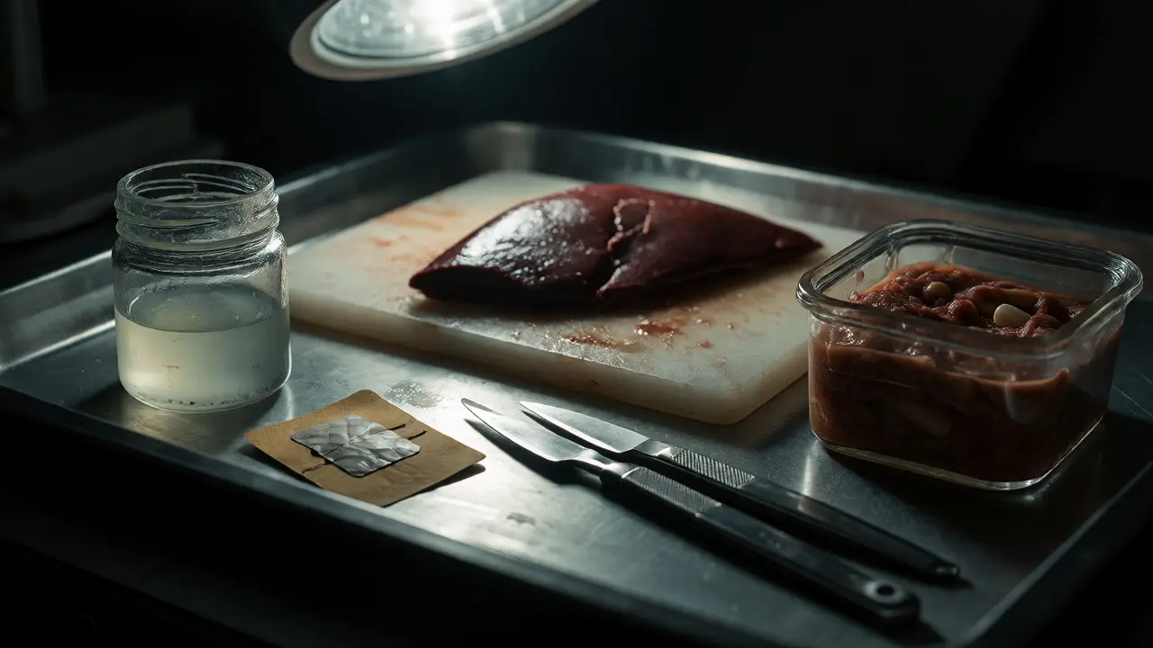
- Gastric Contents: Collect the entire stomach contents if the time of death was within 1-2 hours of ingestion. This helps determine if pills were recently swallowed but not yet absorbed.
- Vitreous Humor: This fluid inside the eye is isolated from blood flow. It’s excellent for estimating time since death and checking electrolytes, but also useful for certain drugs like lithium or ethanol when blood is compromised.
- Liver Tissue: A wedge from the left lobe. Useful for lipophilic drugs (like THC or cocaine metabolites) that accumulate in fat-rich tissues.
Notice I didn’t mention hair? Hair testing is great for long-term history, but it’s rarely collected during a standard emergency autopsy unless chronic abuse is suspected. Save it for specific investigations.
| Specimen | Best For | Key Limitation | Preservative |
|---|---|---|---|
| Femoral Blood | Acute intoxication, legal limits | Postmortem redistribution | Sodium Fluoride |
| Heart Blood | General screening | Highly prone to diffusion from lungs/liver | Sodium Fluoride |
| Urine | Detection window extension | Not quantitative for impairment | Refrigeration |
| Gastric Contents | Recent ingestion evidence | Rapid degradation by acids/enzymes | Refrigeration |
| Vitreous Humor | Time since death, stable analytes | Limited drug panel coverage | None required |
Avoiding Contamination Traps
Contamination is the silent killer of toxicology results. Think about the tools you use. If you used a scalpel that touched formalin-fixed tissue earlier in the day, you might introduce formaldehyde into your sample. Formaldehyde reacts with ammonia and other compounds, creating artifacts that confuse mass spectrometry results.
Use clean, dedicated instruments for each specimen type. Never reuse needles between different body cavities without sterilizing. And here is a big one: external contamination. If the deceased had vomit on their skin, wash the area thoroughly before drawing blood from nearby sites. Vomit contains stomach acid and undigested food, which can alter pH and degrade certain drugs.
Also, consider the environment. Was the body found near a chemical plant? Or did they use topical creams? Transdermal patches (like fentanyl or nicotine) can leave residue on the skin. Document the location of any patches before removing them, and save the patch itself for analysis. The amount of drug remaining in the patch tells you how much was delivered.
Timing Matters More Than You Think
When should you collect samples? Ideally, immediately upon opening the body cavity. Why? Because autolysis begins right away. Enzymes break down proteins and drugs. Bacteria multiply. If you wait until after you’ve removed all the organs and cleaned up, you’ve lost data.
For volatile substances like ethanol, timing is critical. Ethanol can be produced by bacterial fermentation after death (postmortem production). If you don’t have a fluoride tube to stop this process, your "alcohol" result might be fake. Collect blood early, keep it cold, and get it to the lab fast. Refrigeration slows bacterial growth, but freezing preserves integrity longer if there’s a delay.
What if the body is decomposed? Don’t panic. In advanced decomposition, blood may liquefy or clot completely. Switch strategies. Look for solid organs like the psoas muscle or deep thigh muscles. These areas are protected from environmental bacteria and insect activity. Muscle tissue can still yield reliable drug concentrations, though interpretation requires more caution.
Interpreting the Results: Context is King
Once the lab reports come back, you’re not done. A number on a page doesn’t mean anything without context. A therapeutic level of a drug in one person can be lethal in another due to tolerance, age, or comorbidities.
Always correlate toxicology findings with the gross and microscopic pathology. Did you see pulmonary edema? That supports opioid overdose. Did you find coronary artery disease? Then the drug might have been incidental. Did you see petechiae? That points toward mechanical asphyxia, not necessarily poisoning.
Remember the concept of synergistic effects. Alcohol plus benzodiazepines plus opioids creates a respiratory depression cocktail that is far more deadly than any single substance alone. Your report should reflect this complexity. Don’t just list drugs; explain how they interacted with the physical findings.
Practical Tips for the Busy Pathologist
Let’s make this actionable. Here are five rules of thumb I stick to:
- Label everything twice. Name, date, time, site of collection. If you drop a tube, you need to know exactly what it was.
- Take photos. Photograph the stomach contents before discarding them. Show the pill fragments. Visual evidence strengthens your written report.
- Ask about medications. Talk to family or EMS personnel. Knowing the patient was prescribed methadone changes how you interpret a positive morphine test (methadone metabolizes to EDDP, not morphine directly, but cross-reactivity happens).
- Check for embalming. If the body was partially embalmed, formalin will destroy most drug structures. Note this clearly. Some labs can still detect certain metabolites, but many cannot.
- Keep a chain of custody log. Who held the sample? When was it transferred? Missing signatures ruin cases in court.
Finally, build a relationship with your local toxicology lab director. Ask them what they prefer. Do they want whole blood or serum? Do they need 5 mL or 10 mL? Aligning with their protocols reduces rejections and delays.
Why is femoral blood preferred over heart blood for toxicology?
Femoral blood is collected from the leg, far from major organs like the liver and lungs. After death, drugs diffuse out of these organs into the surrounding blood. Heart blood sits right next to the liver, so it often shows artificially high drug concentrations due to this postmortem redistribution. Femoral blood provides a more accurate reflection of circulating drug levels at the time of death.
Can you perform toxicology on a cremated body?
Generally, no. Cremation involves extreme heat that destroys organic molecules, including most drugs and metabolites. However, if bone fragments remain, some specialized labs can attempt analysis on powdered bone, but success rates are low and costs are high. It is best to collect samples before cremation occurs.
What happens if I use the wrong tube for blood collection?
Using a tube without sodium fluoride allows enzymes to break down glucose and potentially alter drug stability. Using a heparin tube might interfere with immunoassays. If you accidentally use the wrong tube, label it clearly as such. The lab may still run tests, but they will note the potential for altered results, which could weaken the legal weight of the finding.
How do you handle vitreous humor collection?
You insert a needle through the sclera (white part of the eye) into the posterior chamber. Withdraw about 1-2 mL per eye. It does not require a preservative because it is naturally sterile and isolated from blood flow. Store it refrigerated. It is particularly useful when blood is unavailable due to decomposition or trauma.
Does embalming fluid affect toxicology results?
Yes, significantly. Formaldehyde in embalming fluid cross-links proteins and degrades many drugs. While some robust compounds like cocaine metabolites or heavy metals might survive, most pharmaceuticals become undetectable. If a body is embalmed, inform the toxicologist immediately. They may adjust their methods or declare the sample unsuitable for certain tests.