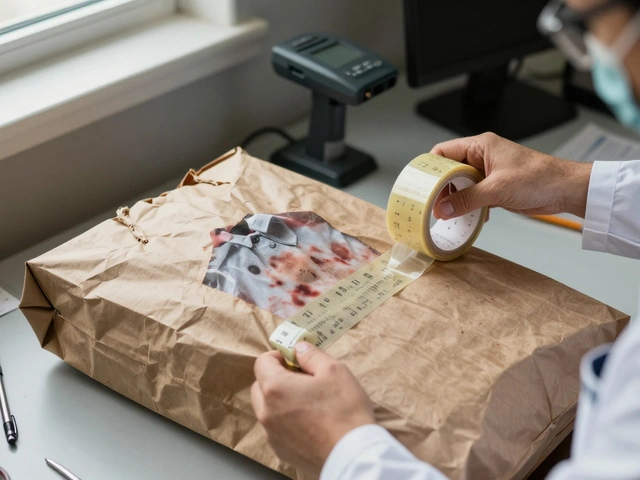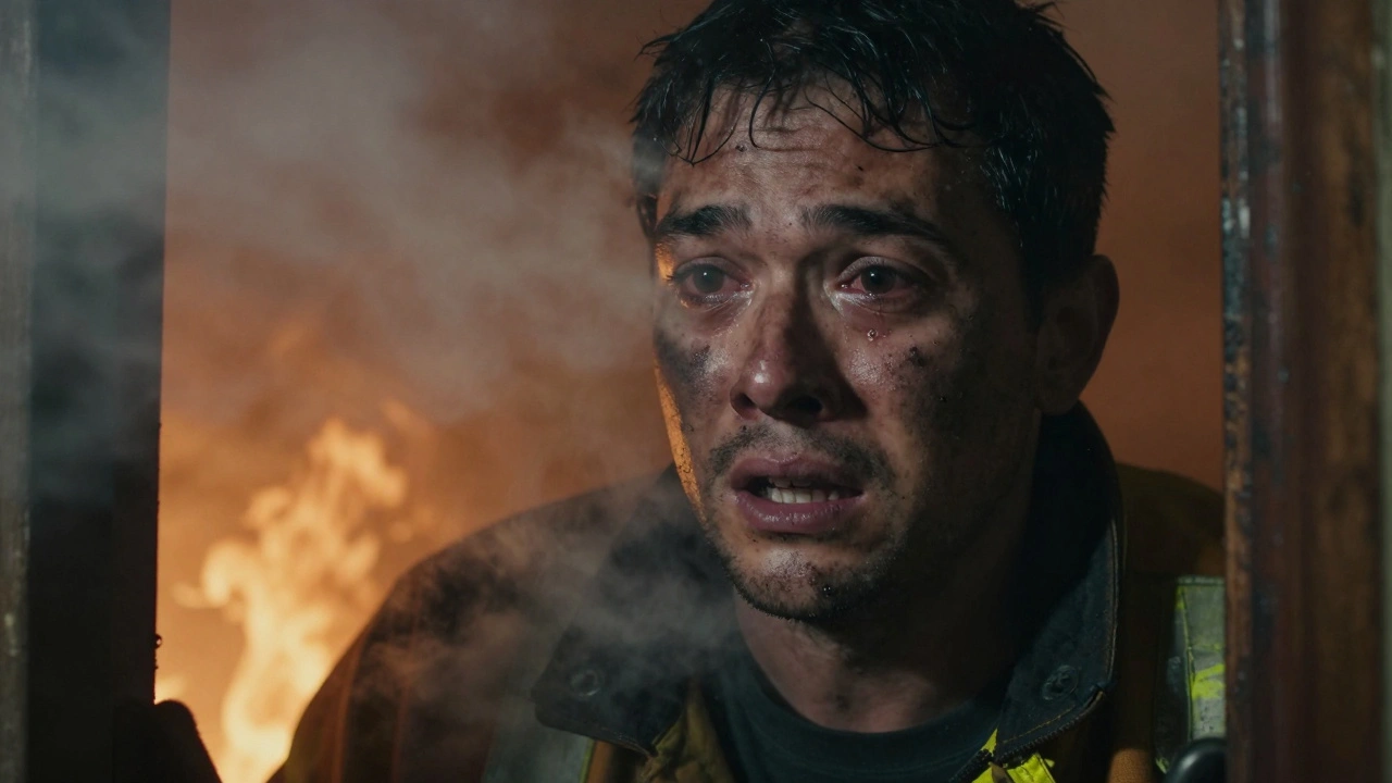
Fires are loud and chaotic, but the damage they cause often happens in silence. You might see the external burns first-the charred skin or the blackened clothing-but the real danger is usually hiding deep inside the breathing tract. When someone survives the initial heat of a blaze, the threat doesn't vanish. It lingers in the throat and lungs, sometimes killing them hours after they leave the burning building. This is thermal airway injury. It is a complex condition that forensics experts and medical teams must evaluate quickly and accurately.
The issue isn’t just about getting burned. It’s about what gets breathed in. We classify these injuries into three distinct categories to understand how they kill patients or determine cause of death in forensic cases. First, you have direct thermal damage hitting the upper airway structures. Next, there’s chemical irritation damaging the lower respiratory tissues. Finally, there is systemic toxicity where poisons like carbon monoxide get absorbed into the blood. Each type plays a role in the overall mortality rate of burn victims.
Key Takeaways
- Thermal airway injury involves tissue damage from smoke, heat, or chemicals during fire exposure.
- Upper airway burns occur above the glottis when air exceeds 150°C, while lower airway damage is mostly chemical.
- Carbon monoxide and hydrogen cyanide are deadly toxins absorbed through the lungs.
- Physical signs include soot in the mouth, hoarseness, and facial burns, but internal injury may require bronchoscopy.
- Diagnosis relies on clinical history, lab tests for carboxyhemoglobin, and direct visualization of the airway.
Understanding the Three Types of Injury
To grasp why this injury is so dangerous, you need to separate the mechanisms. Many people think smoke inhalation is one single problem, but it acts differently depending on where it lands in the body. The upper airway, which includes the face, lips, nose, and throat down to the vocal cords, takes the brunt of the heat. If the air temperature climbs over 150 degrees Celsius, that hot air directly cooks the soft tissue. This creates immediate blistering and swelling right at the top of the tube you breathe through.
This swelling is known as edema. Here is the critical part: the edema doesn’t always show up instantly. A patient might look okay in the ambulance but then swell shut an hour later because the fluid continues to accumulate in the tissues. External burns on the neck or face make this worse because the skin tightens, distorting the anatomy underneath. If the airway narrows too much, the patient can’t pull in enough oxygen.
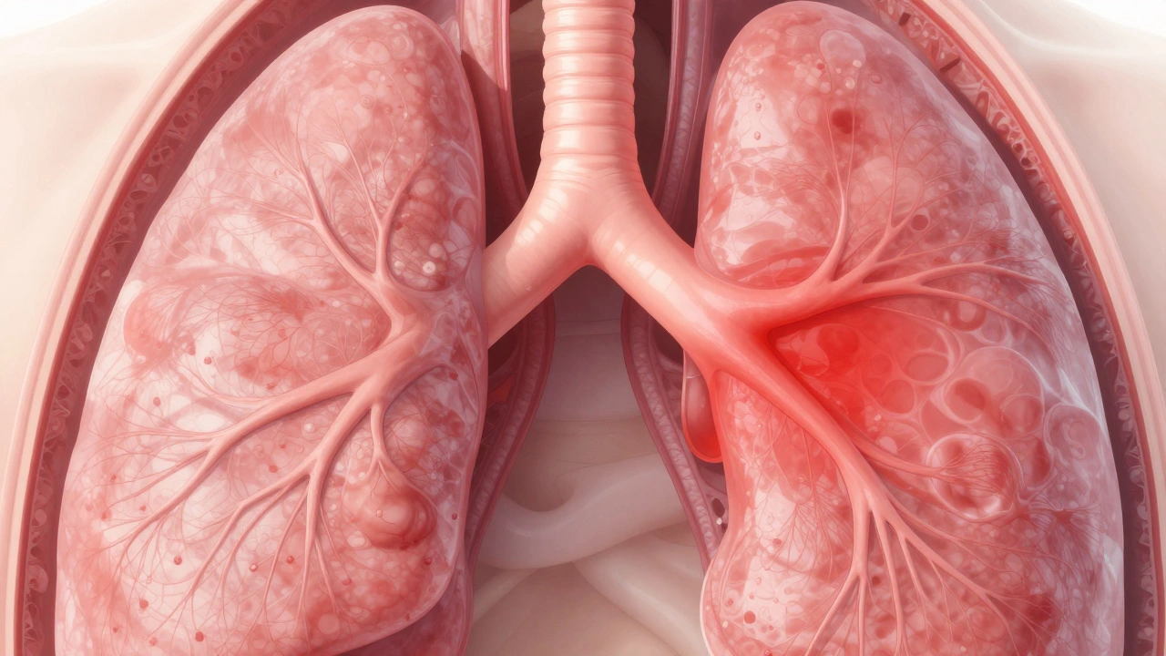
Below the vocal cords, the story changes. The lower airways are cooler by the time the smoke reaches them. Your lungs act as a massive heat exchanger, cooling the incoming air. So, thermal burns deep in the lungs are rare unless you are hit by a steam jet. Instead, the lower lung damage comes from the chemicals in the smoke. These toxins strip away the protective lining of the airways and mess up the tiny hair-like cilia that keep the lungs clean. Without those defenses, bacteria can invade easily, and the fluid balance in the lungs goes haywire, leading to collapse or severe pneumonia later on.
| Feature | Upper Airway Injury | Lower Airway Injury |
|---|---|---|
| Primary Cause | Direct Heat (>150°C) | Chemical Irritants / Toxins |
| Timing | Immediate to 24 Hours (Edema) | Delayed (Hours to Days) |
| Key Symptom | Stridor, Hoarseness, Singed Hair | Coughing, Wheezing, Shortness of Breath |
| Risk Factor | Enclosed Spaces | Prolonged Exposure Duration |
Systemic Poisoning: The Invisible Killers
It is easy to focus on the soot you can see, but the gas molecules are the silent assassins. When fires burn, they produce carbon monoxide (CO) and hydrogen cyanide (HCN). These gases pass through the lungs into the bloodstream, creating a condition called systemic toxicity.
Carbon monoxide binds to hemoglobin in your red blood cells way tighter than oxygen does. This molecule is called carboxyhemoglobin. Once it gets above 15% in the blood, brain cells start dying from lack of oxygen. You won’t see bruises or cuts from this, yet it causes neurological failure or heart attacks in survivors. Similarly, cyanide stops cells from using oxygen even if it gets there. Both need testing. You cannot diagnose CO poisoning just by looking at a bruised face. You need a spectrophotometer to measure the blood levels accurately.
Diagnostic Signs and Physical Examination
When evaluating a suspect death or a live victim, you start with what you can see on the outside. Look at the face and lips. Are they burnt? Is there soot smeared on the lips or inside the mouth? Has the person been coughing so hard they vomited sooty mucus? These are classic red flags.
If you check the neck, listen for sounds. Stridor is a harsh noise made when air struggles to get past swollen tissue. A hoarse voice means the vocal cords are inflamed. If a patient lost consciousness at the scene, their risk is higher because they were likely exposed for longer without protection. However, physical signs alone don't tell the whole story. A person can inhale smoke without having visible soot in the throat, so you have to dig deeper.
The Gold Standard: Bronchoscopy
Sometimes, the eyes aren’t enough. That is where fiberoptic bronchoscopy comes in. This tool is the gold standard for diagnosing true airway injury. Doctors slide a flexible camera down the windpipe. They can see the trachea, the bronchi, and the smaller branches.
What are they looking for? They want to find tracheal soot. More importantly, they look for mucosal injury, erythema (redness), and necrosis (dead tissue). In severe cases, you will see a pseudomembranous cast. Imagine the airway lining peeling off and sticking to itself like a mold. This sloughed-off tissue blocks the tube completely. Finding this early helps predict who will develop Acute Respiratory Distress Syndrome (ARDS). While the camera can miss some disease in the tiny air sacs (parenchyma), it tells you exactly what the big pipes look like.
If the airway looks threatened, doctors might perform intubation via the awake fiberoptic technique. Trying to intubate blindly when the throat is swelling is risky because you might tear the tissue further. Visualization keeps it safe. Even though bronchoscopy doesn’t replace the need for monitoring, it provides the evidence needed to justify aggressive care.

Laboratory Markers and Imaging
Blood work supports what the eyes miss. You need to run a Complete Blood Count (CBC) and a basic metabolic panel. Crucially, you measure lactate and venous blood gas (VBG). High lactate suggests the body is starving for oxygen. Chest X-rays are tricky because they often look normal in the beginning. The fluid accumulation takes time to settle. However, getting a baseline image matters. If the x-ray is clear today but cloudy tomorrow, you know the inflammation has progressed.
We also check methemoglobin levels alongside carboxyhemoglobin. These numbers give us a snapshot of the toxic load. Remember, finding soot in secretions confirms smoke exposure, but it doesn’t automatically mean the injury is fatal. You have to combine the lab results with the timeline of events at the scene.
Timeline of Complications
You need to respect the clock. Airway swelling peaks around 24 hours after the incident. A patient breathing fine at midnight might be unable to breathe at dawn. The combination of internal swelling and external skin burns makes managing the airway incredibly difficult. Fluid resuscitation, which treats the burns, actually pumps more water into the swollen throat, narrowing the space further.
This dynamic progression is why repeat assessments are mandatory. You cannot just admit a patient once and leave. The diagnosis of lower airway damage depends heavily on the clinical course rather than the initial findings. Wheezing or rhonchi (rattling sounds) might appear days later as the airways spasm. Keeping watch is just as important as the initial test.
Summary of Evaluation Protocols
Evaluation isn’t just one test. It is a checklist built on layers of evidence. You need a history of enclosed space fire, visual confirmation of burns or soot, clinical signs of distress, and lab confirmation of toxins. Missing one piece doesn’t rule out injury, but gathering them all solidifies the case. In forensics, determining if death was caused by the fire or the subsequent airway closure impacts legal outcomes significantly.
Can soot in the throat confirm thermal injury?
Not necessarily. Soot indicates smoke exposure, but it does not confirm the severity of the injury. Severe thermal damage often shows swelling, blistering, and ulceration beyond just soot deposits. You must evaluate tissue health visually or via bronchoscopy.
How soon does airway swelling peak?
Swelling typically progresses significantly over the first 24 hours following the injury. Clinical evaluations must be repeated frequently during this window to detect rapid changes in airway patency.
Is bronchoscopy necessary for every smoke inhalation patient?
It is not required for every minor case, but it is the gold standard for suspected severe injury. If there are facial burns, hoarseness, or a closed-space fire history, bronchoscopy is highly recommended to grade damage severity.
What blood test detects carbon monoxide poisoning?
A co-oximetry test or spectrophotometry is used to measure carboxyhemoglobin (COHb) levels in the blood. Levels exceeding 15% indicate significant toxicity requiring intervention.
Why is lower airway damage mostly chemical?
The air cools rapidly as it passes the trachea. Therefore, the lower airway rarely suffers thermal burns. Damage occurs due to inhaled toxic chemicals in smoke causing epithelial cell death and increased capillary permeability.
Understanding the mechanics of thermal airway injury saves lives and clarifies forensic findings. By recognizing the triad of thermal burns, chemical irritation, and systemic poison, medical teams can intervene before the airway closes permanently. Always remember that the most dangerous part of a fire might not be the flame itself, but what follows the breath.