When a person dies from lack of oxygen, the body doesn't always leave obvious clues. There might be no bruises on the neck. There might be no broken bones. Yet, someone has been killed by strangulation or asphyxia. For forensic pathologists, determining exactly how this happened is one of the most complex challenges in medicine. It requires piecing together microscopic tissue damage, scene evidence, and medical history to distinguish between suicide, homicide, and accident.
The difference between strangulation, which involves external pressure on the neck, and broader forms of asphyxia, such as suffocation or choking, is critical for legal proceedings. A misclassification can mean the difference between justice served and a killer walking free. This guide breaks down how experts determine the cause and manner of death in these cases, moving beyond myths about "broken necks" to the reality of soft tissue trauma and physiological collapse.
The Physiology of Death by Neck Compression
Many people believe that strangulation kills by cutting off air. While blocking the windpipe (trachea) does lead to death, it is actually the slowest mechanism. The human brain can survive without oxygen for several minutes before permanent damage occurs. In contrast, compressing the blood vessels in the neck leads to unconsciousness in seconds and death within minutes.
Forensic literature identifies four primary mechanisms at play during neck compression:
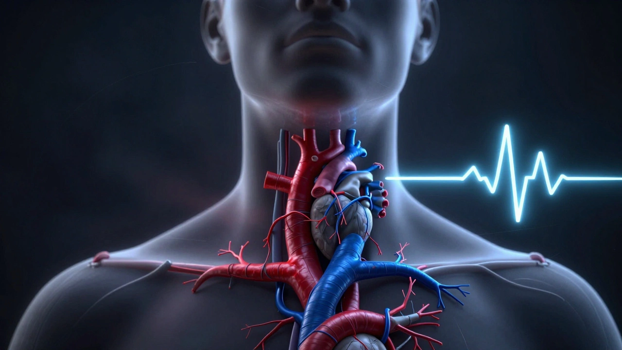
- Carotid Artery Occlusion: Pressure on the major arteries supplying the brain cuts off oxygenated blood. Unconsciousness typically occurs within 10 seconds. This is often the fastest route to death.
- Jugular Vein Obstruction: Compressing the veins prevents blood from leaving the brain. This causes congestion, swelling, and eventually stops breathing. It takes longer than arterial occlusion but is still rapid.
- Airway Blockage: Crushing the larynx or trachea stops airflow. This produces classic signs of asphyxia like blue skin (cyanosis), but it requires significantly more force and time than vascular compression.
- Reflex Cardiac Arrest: Stimulation of the carotid sinus nerve ganglion can trigger a fatal heart rhythm disturbance almost instantly, even if blood flow isn't completely stopped.
In real-world scenarios, these mechanisms rarely happen in isolation. A ligature or hands usually compress multiple structures simultaneously. Understanding this physiology helps pathologists look for specific types of internal damage rather than just external bruising.
Types of Strangulation and Their Signatures
Not all neck compression looks the same. Forensic pathologists categorize deaths into three main types based on the method used. Each type leaves distinct traces, though there is significant overlap in internal injuries.
| Type | Mechanism | Typical Manner of Death | Key Forensic Indicators |
|---|---|---|---|
| Hanging | Body weight suspends the victim via a ligature. | Mostly Suicide; some Homicide/Accident. | High ligature mark, often oblique; minimal struggle signs; fracture of hyoid/larynx common in older adults. |
| Ligature Strangulation | External force tightens a ligature around the neck (e.g., rope, belt). | Mostly Homicide; rare Suicide. | Horizontal ligature mark; extensive soft tissue hemorrhage; signs of struggle at scene. |
| Manual Strangulation | Hands, arms, or objects applied directly to the neck. | Almost exclusively Homicide. | Fingerprints or fingernail marks; deep muscle hemorrhages; frequent association with domestic violence. |
It is crucial to note that hanging is defined by the use of the victim's own body weight to tighten the ligature. If another person applies the force, it is classified as ligature strangulation, regardless of whether the body is suspended. This distinction is vital for determining the manner of death.
The Autopsy: Looking Beyond the Skin
The most dangerous myth in forensic pathology is that strangulation always leaves visible bruises. In many cases, especially manual strangulation, the external skin may appear pristine. The real evidence lies beneath the surface.
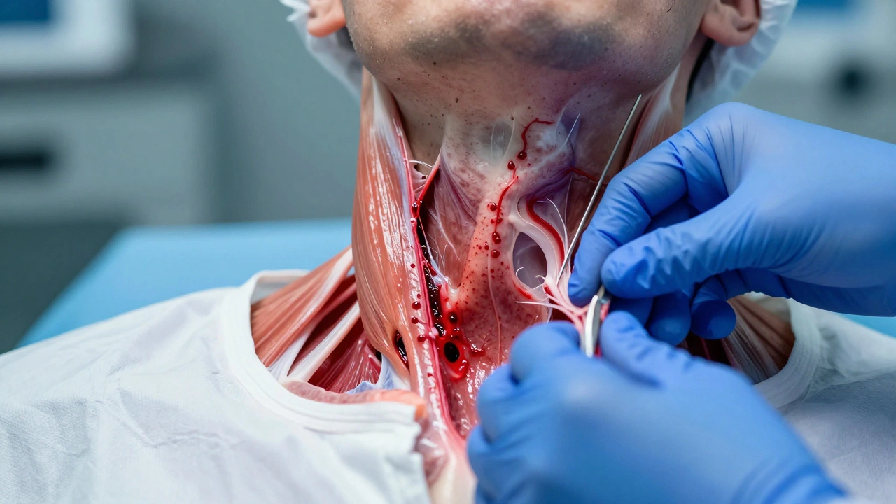
A thorough autopsy for suspected strangulation involves a meticulous dissection of the neck. Pathologists remove the tongue, larynx, and hyoid bone as a single block to preserve spatial relationships. They then slice through the layers of muscle-specifically the sternohyoid and sternothyroid muscles-to look for tiny hemorrhages. These small bleeds are caused by the rupture of capillaries under pressure and are strong indicators of ante-mortem trauma.
Bone fractures are less common than pop culture suggests. Studies show that hyoid bone fractures occur in no more than one-third of fatal strangulations. They are more likely in older individuals whose bones have become brittle. Conversely, younger victims often suffer severe soft tissue damage without any bone breaks. Therefore, the absence of a fractured hyoid does not rule out strangulation.
Petechiae: The Misunderstood Marker
You will often hear about petechiae-tiny, pinpoint red spots found in the eyes, eyelids, or face-in discussions of asphyxia. These spots result from ruptured capillaries due to increased pressure in the head's venous system.
While petechiae are found in up to 95% of manual strangulation cases, they are not exclusive to it. They can also appear in:
- Sudden cardiac arrest
- Coughing or vomiting violently
- Drowning
- SIDS (Sudden Infant Death Syndrome)
- Post-mortem positioning (if the head is lower than the body after death)
Because of this lack of specificity, pathologists never rely on petechiae alone to prove strangulation. Instead, they look for the combination of petechiae, internal neck muscle hemorrhages, and a consistent scene investigation. When all three align, the conclusion becomes robust.
Scene Investigation and Manner of Death
Determining the cause of death (strangulation) is different from determining the manner of death (homicide, suicide, accident, undetermined). The scene provides the context that the autopsy cannot.
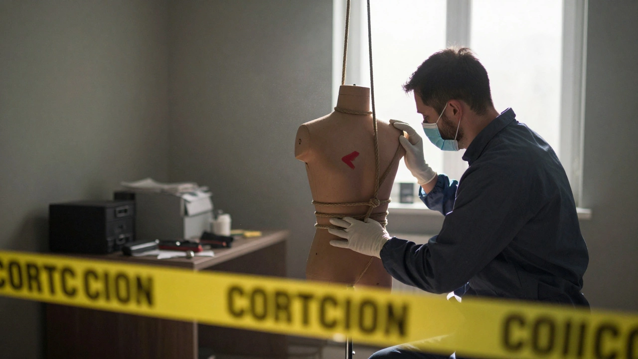
In cases of hanging, investigators look for the position of the knot, the height of the suspension point, and the presence of ligature marks. A high, oblique mark often suggests true hanging (suicide), while a horizontal mark across the front of the neck suggests ligature strangulation (homicide). However, scenes can be staged. A murderer might hang a victim to make it look like suicide. This is why forensic teams correlate scene findings with injury patterns. Homicidal strangulation often presents with additional defensive wounds, bruising on other parts of the body, or signs of a struggle, whereas suicidal hangings typically lack these extraneous injuries.
In infant deaths, the line between SIDS and accidental asphyxia (such as overlaying by a parent or entrapment in bedding) is thin. The CDC’s updated guidelines emphasize detailed documentation of the sleep environment. Without a thorough scene inspection, an accidental suffocation could be misclassified as natural death, missing a preventable tragedy.
Complications: Resuscitation and Organ Donation
Modern medicine sometimes obscures forensic evidence. Aggressive CPR can cause rib fractures, lung contusions, and even petechiae, mimicking the signs of asphyxia. More critically, organ procurement procedures can destroy key evidence. Harvesting the heart often involves cutting through the chest and neck tissues, potentially obliterating subtle hemorrhages in the strap muscles or vocal cords.
Pathologists must carefully document any prior medical interventions. If a victim was resuscitated or donated organs, the pathologist relies more heavily on imaging data collected before those procedures and on the circumstantial evidence from the crime scene. This highlights the need for immediate preservation of evidence before hospital interventions alter the body.
Advanced Imaging and Future Techniques
Traditional autopsies are being supplemented by advanced imaging. Post-mortem computed tomography (PMCT) is excellent for detecting fractures of the hyoid and laryngeal cartilages that might be missed during visual inspection. It provides a 3D map of bony injuries, which is invaluable for courtroom presentations.
However, MRI and CT scans have limitations. They may miss small intramuscular hemorrhages or subtle vocal cord bleeding that only physical dissection can reveal. Currently, no biochemical test can definitively distinguish between ante-mortem and post-mortem neck trauma, although research continues. For now, the combination of expert dissection, radiological imaging, and rigorous scene analysis remains the gold standard.
Can you die from strangulation without any external marks?
Yes. Manual strangulation can cause death by compressing blood vessels or triggering a reflex cardiac arrest without leaving visible bruises on the skin. Internal hemorrhages in the neck muscles are often the only physical evidence, requiring a detailed autopsy to detect.
What is the difference between hanging and ligature strangulation?
Hanging uses the victim's own body weight to tighten the ligature, often resulting in a diagonal mark. Ligature strangulation involves an external force tightening the ligature, usually resulting in a horizontal mark. Hanging is frequently associated with suicide, while ligature strangulation is typically homicidal.
Are petechiae proof of strangulation?
No. Petechiae (pinpoint hemorrhages in the eyes or face) are common in strangulation but also occur in heart attacks, coughing fits, drowning, and SIDS. They must be interpreted alongside neck injuries and scene evidence to confirm strangulation.
How do pathologists distinguish between suicide and homicide in neck compression cases?
Pathologists integrate autopsy findings with scene investigation. Homicides often show signs of struggle, defensive wounds, or extensive soft tissue trauma inconsistent with self-infliction. Suicides typically lack these additional injuries and may show specific ligature patterns indicative of self-suspension.
Does CPR interfere with determining the cause of death?
Yes. CPR can cause rib fractures, lung damage, and petechiae that mimic asphyxia. Organ donation procedures can also destroy subtle neck injuries. Pathologists must account for these iatrogenic changes when analyzing the body.