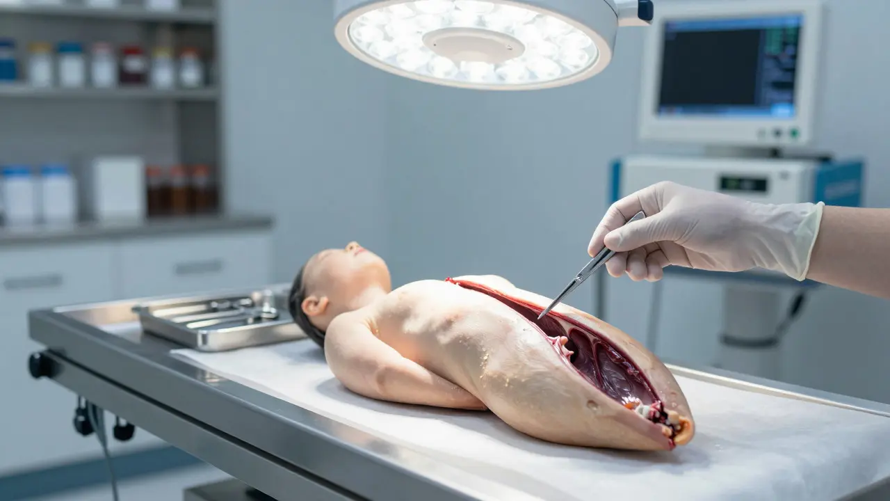
When a body arrives at the morgue, the clock is ticking. The most critical evidence isn't always under a microscope; it's often visible to the naked eye during the initial organ examination. This systematic process of analyzing and dissecting internal organs is the backbone of determining cause and manner of death. It’s not just about cutting tissue; it’s about reading the story written in the organ's structure, weight, and color.
For forensic pathologists, this procedure bridges the gap between external injury and internal physiological failure. Whether the case involves trauma, poisoning, or sudden natural death, the organs hold the final answer. Understanding how this analysis works reveals why some autopsies take hours while others require weeks of lab work.
The Core Purpose of Systematic Organ Analysis
Systematic organ analysis is a standardized protocol used by forensic pathologists to inspect, weigh, and section internal organs to identify pathological changes. Unlike clinical autopsies which might focus on specific diseases, forensic examinations must rule out everything from homicide to rare genetic disorders. The goal is consistency. If two different pathologists examine the same heart, they should find the same abnormalities. That standardization relies on strict protocols established by bodies like the American Board of Forensic Pathology.
This process serves three main jobs:
- Cause of Death Determination: Identifying if an organ failed due to disease (like a heart attack) or external force (like blunt impact).
- Manner of Death Classification: Distinguishing between accident, suicide, homicide, or undetermined cases based on physical evidence.
- Evidence Preservation: Securing tissues for toxicology, histology, and DNA testing before decomposition alters the scene.
Step-by-Step: How the Dissection Unfolds
The actual dissection follows a logical sequence, usually starting with the head and moving down to the pelvis. This order prevents cross-contamination and ensures no area is missed. Here is how a typical full-body autopsy proceeds regarding internal organs:
- External Inspection & Y-Incision: Before touching organs, the skin is cut in a 'Y' shape from shoulder to shoulder and down to the pubic bone. This exposes the chest and abdomen.
- Thoracic Cavity Access: The rib cage is cut open. The lungs are examined first for bruising, aspiration, or collapse. Then, the heart is removed along with the great vessels.
- Abdominal Cavity Access: The abdominal wall is opened. The stomach, intestines, liver, spleen, and kidneys are inspected. The peritoneal cavity is checked for fluid (blood or pus).
- Pelvic Cavity Access: The bladder, reproductive organs, and lower intestines are examined.
Each step requires documentation. Photos are taken before, during, and after removal. Weights are recorded immediately. A normal adult human heart weighs between 250g and 350g. If it’s 400g, that’s hypertrophy-a sign of long-term high blood pressure or valve disease. These numbers matter more than you might think.
Gross Anatomy vs. Microscopic Analysis
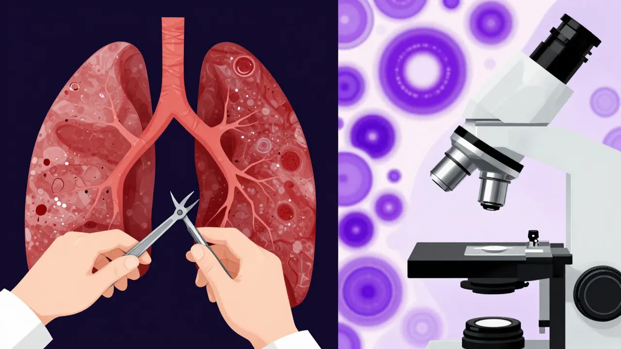
Many people assume the autopsy is over once the organs are out. In reality, that’s only half the battle. Gross examination uses the eyes, hands, and knives. Microscopic analysis uses slides and stains. They complement each other perfectly.
| Feature | Gross Examination | Microscopic Analysis |
|---|---|---|
| Tools Used | Surgical scalpel, scissors, scales | Light microscope, hematoxylin-eosin stains |
| Time Frame | During autopsy (hours) | Laboratory phase (days to weeks) |
| Detects | Friactures, masses, discoloration, size changes | Cellular damage, early infection, micro-fractures, drug deposits |
| Example Finding | Enlarged liver (hepatomegaly) | Steatosis (fatty liver) or cirrhosis patterns |
Consider a case of suspected carbon monoxide poisoning. Grossly, the victim’s lips and skin might look cherry-red. But the definitive proof often comes from microscopic analysis of the lung tissue or even post-mortem chemistry. Conversely, a subtle brain hemorrhage might be invisible to the eye but obvious under a microscope as extravasated red blood cells. Missing either layer risks misdiagnosis.
Key Organs and What Pathologists Look For
Not all organs get the same level of scrutiny. Some are high-yield for common causes of death. Let’s break down the big four: heart, lungs, liver, and brain.
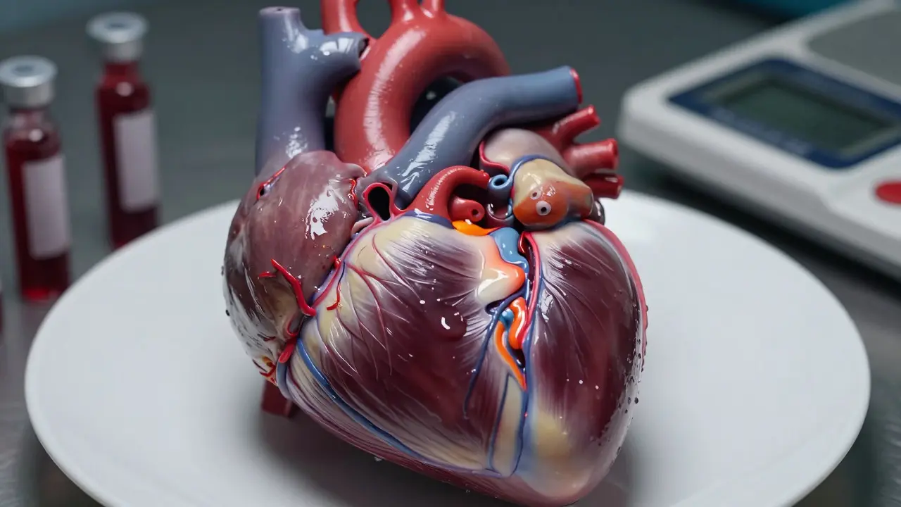
The Heart: The Engine Room
Cardiac examination is the detailed inspection of the heart muscle, valves, and coronary arteries to determine cardiac function and pathology. Coronary artery disease remains the leading cause of natural death globally. During dissection, the pathologist sections the coronary arteries perpendicular to their length. Why? To see the lumen (inside). A 70% blockage looks different from a 90% blockage. They also check for thrombus (clot) formation. Was the clot fresh (red and soft) or old (yellow and hard)? Fresh means it likely caused the death. Old means it was present for weeks.
The Lungs: The Gas Exchange Hub
Lung examination focuses on volume, texture, and presence of foreign material. In drowning cases, the lungs might contain foam mixed with water. In smoke inhalation, they may be blackened and edematous. Aspiration pneumonia-where vomit or food enters the airway-is another key finding. The pathologist cuts the lung into wedges to check for consolidation (solidification) or abscesses.
The Liver: The Metabolic Filter
The liver processes drugs, toxins, and nutrients. Its appearance tells a lot about lifestyle and acute events. Fatty liver (steatosis) appears yellow and waxy. Cirrhosis makes the surface bumpy and nodular. In blunt trauma, the liver is prone to rupture because it’s large and fixed in place. A torn capsule releases blood into the abdomen, which is easily seen during the opening of the abdominal cavity.
The Brain: The Control Center
Brain examination is delicate. The skull is opened, and the brain is lifted out. The meninges (coverings) are checked for bleeding. Subdural hematoma (bleeding between the brain and skull) is a classic sign of acceleration-deceleration injuries, common in falls or assaults. The brain tissue itself is sliced horizontally to reveal any contusions (bruises) or herniation (shifting of brain parts), which indicates raised intracranial pressure.
Common Pitfalls in Organ Dissection
Even experienced pathologists make mistakes. Knowing where errors happen helps improve accuracy.
- Artifacts from Handling: Cutting too roughly can create tears that look like traumatic injuries. Using proper technique minimizes this.
- Decomposition Changes: In warm weather, gas production can mimic internal injuries. Green discoloration of the abdomen can be mistaken for bruising. Context is key.
- Missing Small Lesions: A small pulmonary embolism (blood clot in the lung artery) can be hidden in the peripheral branches. Thorough sectioning of the pulmonary trunk is essential.
- Assuming Natural Causes Too Early: If a patient has known heart disease, pathologists might stop looking for trauma. Always rule out homicide before confirming natural death.
The Role of Toxicology in Organ Analysis
Organ examination doesn’t end with histology. Tissues are sent to the toxicology lab. Blood, urine, vitreous humor (eye fluid), and bile are tested for drugs and poisons. Sometimes, the liver or kidney is specifically sampled for chronic exposure markers. For example, heavy metal poisoning leaves distinct deposits in renal tubules. Without these samples, a case of arsenic poisoning might be missed entirely if the symptoms were non-specific.
Documentation: The Legal Backbone
If it isn’t written down, it didn’t happen. In court, the autopsy report is the primary witness. Photos must be labeled with scale bars. Descriptions must be precise. Instead of saying "the liver looked bad," write "liver weighed 1800g, surface showed coarse nodularity consistent with cirrhosis." Specificity protects the pathologist and informs the jury. Digital tools now allow 3D reconstruction of organs, but the traditional knife-and-eye method remains the gold standard for immediate assessment.
Frequently Asked Questions
How long does a full organ examination take?
A standard full-body autopsy typically takes 2 to 4 hours for the gross examination. However, the total time to complete the case, including waiting for microscopic results and toxicology reports, can range from 2 to 6 weeks.
Do all organs need to be removed during an autopsy?
In a full autopsy, yes, major organs are removed for inspection. In a limited autopsy, only specific organs relevant to the case are examined. The decision depends on the scope of the investigation and legal requirements.
What is the difference between cause and manner of death?
Cause of death is the medical condition or injury that started the chain of events leading to death (e.g., myocardial infarction). Manner of death is the classification of how it happened: natural, accident, homicide, suicide, or undetermined.
Can organ examination detect drug use?
Yes, but indirectly. While visual inspection won't show drugs, tissue samples are sent for toxicology screening. Certain drugs can cause specific organ changes, like liver damage from acetaminophen overdose, which can be seen microscopically.
Why is weighing organs important?
Organ weights provide objective data on health status. Significant deviation from normal ranges indicates pathology. For example, an enlarged heart suggests hypertension, while a shrunken kidney suggests chronic disease.