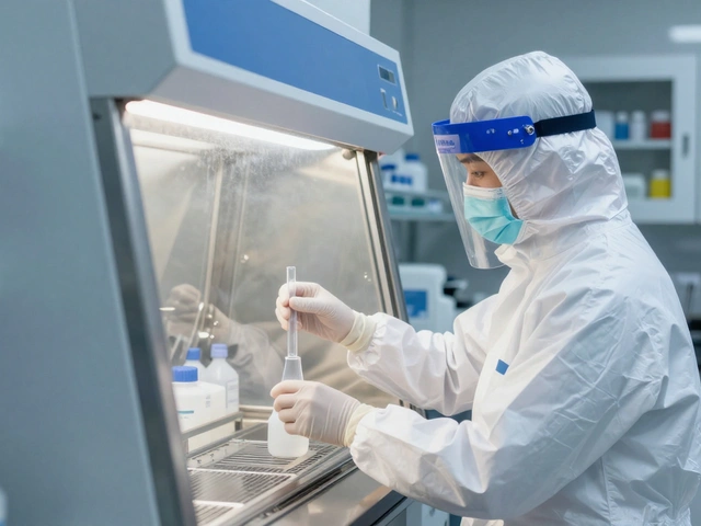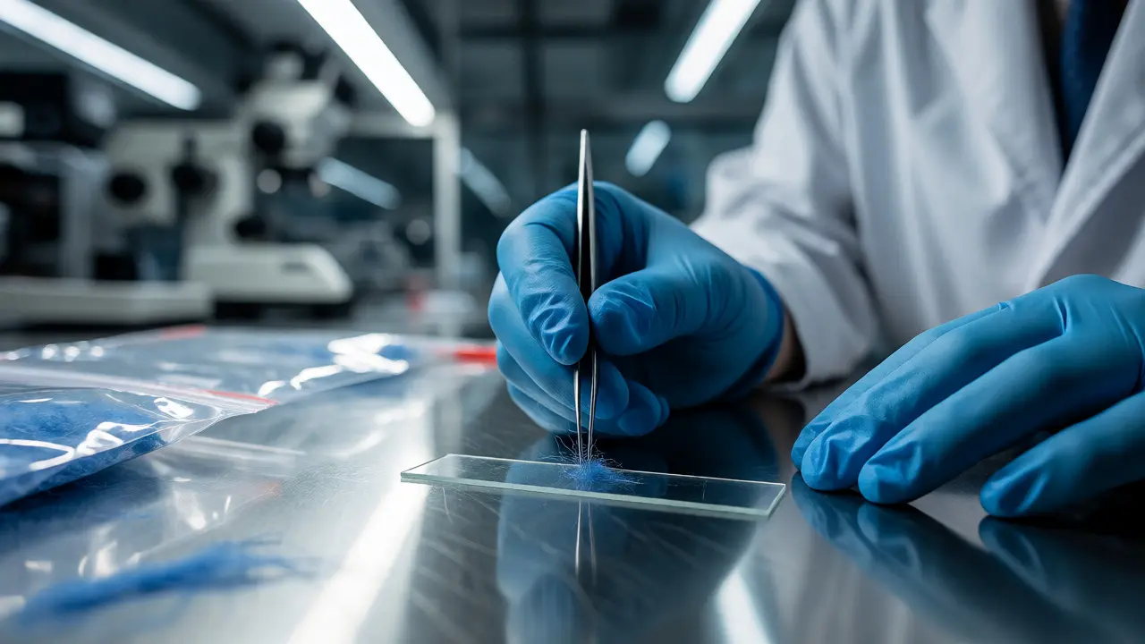
Imagine a single blue fiber found on a suspect’s jacket matches the exact weave of a victim’s carpet. To the naked eye, it’s just lint. To a forensic examiner, it’s a smoking gun. But how do you prove that two microscopic objects are identical when they look like nothing more than specks of dust? The answer lies in microscopy is the primary analytical technique used in trace evidence examination to identify and compare physical materials such as fibers, hairs, glass, paint, and soil. It transforms invisible clues into courtroom testimony.
You might think DNA solves everything these days, but trace evidence remains critical because it links people to places or things in ways biology often can’t. You don’t need blood or saliva to place someone at a scene; you just need them to brush against a surface. That transfer leaves behind trace evidence, which consists of small fragments of matter transferred between persons, objects, or locations during contact. Microscopy is the tool that makes sense of this debris. Whether you’re a student, an investigator, or just curious about how CSI actually works, understanding the microscope’s role changes how you view a crime scene.
The Core Principle: Comparison Over Identification
Most people misunderstand what forensic scientists do with microscopes. They aren’t usually trying to say, "This fiber is definitely nylon." While identification matters, the real magic happens in comparison. The question isn't always "What is this?" but rather "Does this match that?"
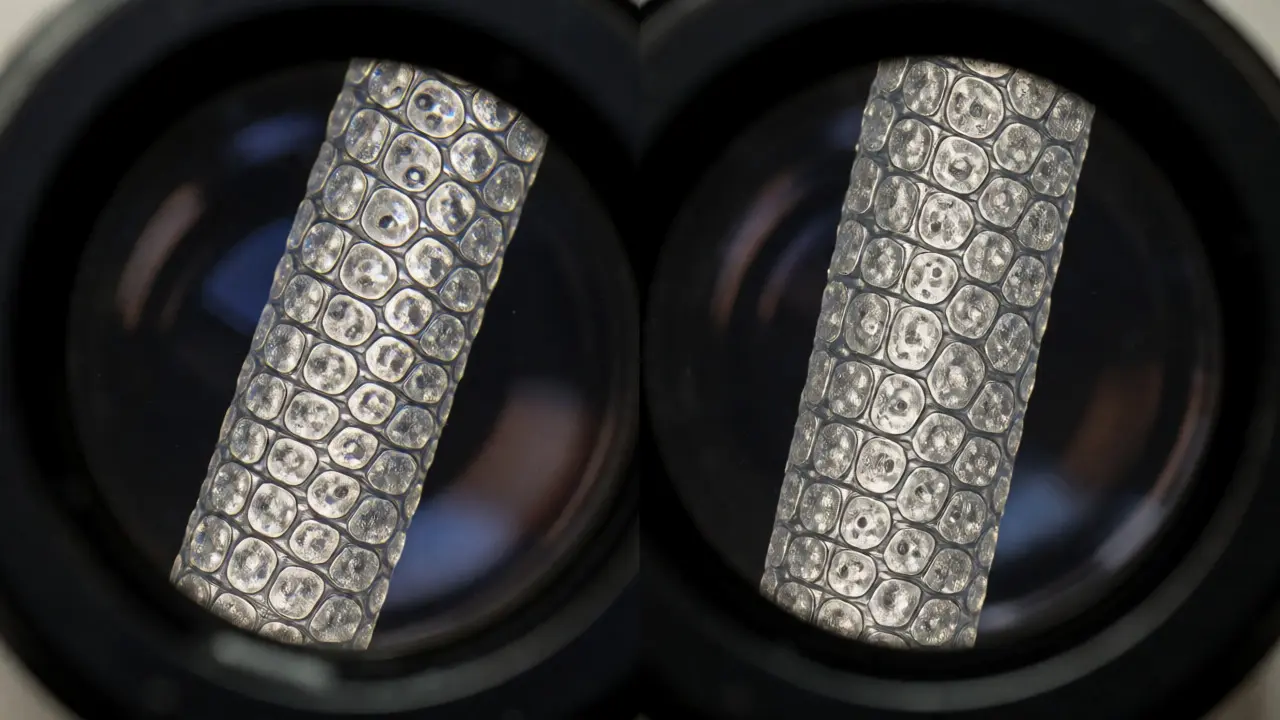
This is where the comparison microscope becomes essential. It is a specialized optical instrument that allows an examiner to view two specimens side-by-side through a single eyepiece. Unlike standard lab microscopes, this device lets you see the suspect’s fiber and the victim’s fiber simultaneously. If the striations on a hair shaft align perfectly, or if the refractive index of a glass shard matches a broken window pane exactly, you have a strong link.
Think of it like matching puzzle pieces. You don’t need to know who made the puzzle; you just need to prove the edges fit. In court, an examiner might testify that the evidence is "consistent with" the source, not necessarily unique to it. Why? Because millions of people wear the same brand of jeans. But if the microscopic wear patterns on those jeans match the unique tear pattern on the victim’s shirt, that’s powerful context.
Types of Microscopy Used in Forensics
Not all microscopes are created equal. Different types of trace evidence require different visual approaches. Using the wrong one could mean missing critical details entirely.
- Stereomicroscopes: These are your workhorses for initial screening. They provide low magnification (usually up to 40x) but offer great depth of field. Examiners use them to sift through vacuum sweepings from a car seat or clothing to find potential candidates for further analysis. You wouldn’t use a high-power lens here because you’d lose track of the sample instantly.
- Compound Light Microscopes: Once you’ve isolated a candidate, you move to higher magnification (up to 1000x). This helps examine cellular structures in hair or pigment particles in paint. However, regular light microscopy has limits. Many synthetic fibers look identical under visible light.
- Polarized Light Microscopy (PLM): This is the gold standard for identifying unknown crystalline materials. By passing light through filters, PLM reveals optical properties like birefringence. Glass, minerals, and some plastics glow or change color in specific ways under polarized light. For example, quartz and calcite behave differently, allowing examiners to distinguish between natural soils and manufactured fillers.
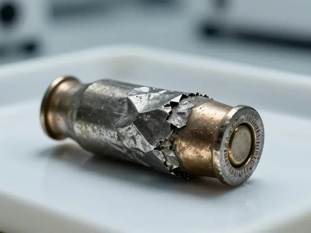
- Scanning Electron Microscope (SEM): When optical methods hit their limit, SEM takes over. It uses electrons instead of light, providing massive depth of field and resolution down to nanometers. SEM coupled with Energy Dispersive X-ray Spectroscopy (EDS) doesn’t just show you what something looks like; it tells you what elements it contains. A gunshot residue particle isn’t just a sphere; it’s a lead-antimony-barium complex confirmed by elemental spikes.
Analyzing Specific Trace Materials
Each type of trace evidence behaves differently under the lens. Here is how examiners tackle the most common categories.
Hair Analysis
Human hair is composed of three layers: the cuticle (outer scale), the cortex (middle pigment layer), and sometimes the medulla (center core). Under a microscope, the shape of the scales, the distribution of pigment granules, and the width of the medulla help determine race and body location. More importantly, comparing two hairs side-by-side can reveal if they came from the same person. If the cuticle patterns are identical and the pigment distribution matches, the probability of coincidence drops significantly. Note that nuclear DNA extraction destroys the hair root, so morphological comparison via microscopy is often done first to preserve the sample for genetic testing later.
Fiber Examination
Fibers are ubiquitous. We shed them constantly. But not all fibers are equal. Natural fibers like cotton and wool have distinct cross-sectional shapes-cotton looks twisted like a ribbon, while wool has overlapping scales similar to human hair. Synthetic fibers like polyester or nylon are extruded, meaning they are forced through tiny holes, resulting in uniform cylindrical shapes with specific cross-sections (round, trilobal, dogbone).
Examiners use Polarized Light Microscopy to measure the refractive index and birefringence of fibers, which are intrinsic physical properties independent of dye. Dyes fade or vary by batch, but the polymer structure doesn’t lie. If a suspect’s jacket fiber has the same refractive index as the victim’s blanket fiber, that’s a solid link.
Glass Fragments
Glass breaks in predictable ways. Radial fractures spread outward from the impact point, while concentric fractures form rings around it. By examining the edge characteristics under a stereoscope, investigators can determine the direction of force. Was the window broken from inside or outside?
For chemical matching, we rely on the Refractive Index (RI). Every piece of glass has a unique RI based on its chemical composition. An examiner immerses a tiny glass fragment in oils with known RIs until the fragment seems to disappear (matching the oil’s RI). If the suspect’s shoe sole contains a glass shard with the exact same RI as the crime scene window, the connection is strong. Modern labs use automated systems to measure RI to four decimal places, making false positives rare.
The Workflow: From Scene to Courtroom
How does a messy pile of debris become evidence? The process is rigorous to prevent contamination and bias.
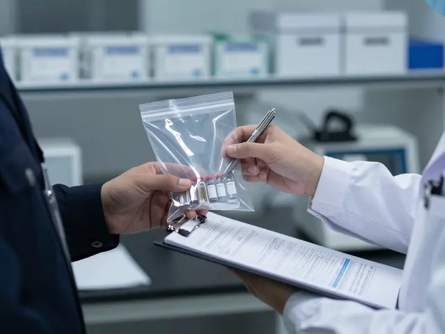
- Collection: Investigators use tape lifts, vacuums, or tweezers to gather samples. Chain of custody starts immediately. Each item is bagged separately to avoid cross-contamination.
- Screening: Back in the lab, the technician uses a stereomicroscope to sort the material. They separate obvious non-evidence (like food crumbs) from potential trace materials.
- Mounting: Candidate items are mounted on slides using mounting media. This preserves them and prepares them for high-magnification viewing.
- Analysis: The examiner runs the battery of tests described above-optical, chemical, and instrumental.
- Reporting: The findings are documented with photographs taken through the microscope. These images are crucial for jury comprehension. A photo of two matching hair cuticles speaks louder than a paragraph of text.
Limitations and Misconceptions
Despite its power, microscopy isn’t infallible. The biggest misconception is the idea of absolute certainty. Forensic examiners rarely say "match." They say "consistent with." Why? Because mass production means many products share identical microscopic features. A generic white cotton T-shirt sold at Walmart looks identical to another one bought at Target. Finding one on the other proves contact, but not identity.
Another limitation is subjectivity. Two experienced examiners might disagree on whether a paint chip’s layer sequence is "similar" or "identical." To combat this, labs follow strict standards set by organizations like the American Society for Testing and Materials (ASTM). Blind verification, where a second examiner analyzes the evidence without knowing the first examiner’s conclusion, reduces bias.
| Technique | Magnification | Primary Use Case | Key Advantage |
|---|---|---|---|
| Stereomicroscope | Low (10x-40x) | Initial sorting, gross morphology | Large depth of field, easy handling |
| Comparison Microscope | Medium (40x-100x) | Side-by-side matching (hair, ballistics) | Direct visual alignment of features |
| Polarized Light (PLM) | High (100x-400x) | Crystalline solids, fibers, glass | Identifies optical properties (RI, birefringence) |
| Scanning Electron (SEM) | Very High (1000x+) | Nanoparticles, gunshot residue, complex surfaces | Elemental analysis combined with imaging |
Why Microscopy Still Matters in the Age of DNA
DNA profiling revolutionized forensics, but it didn’t replace microscopy. DNA requires biological material-cells with nuclei. Hair shafts often lack sufficient DNA. Fibers, glass, and paint contain no DNA at all. Yet, these materials transfer easily and persist longer than biological fluids.
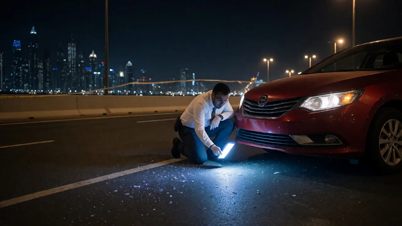
Consider a hit-and-run case. There’s no blood, no skin cells. Just a few flakes of red paint on the bumper and a shard of headlight glass on the road. Without microscopy, there’s no case. With it, the paint’s layered structure and chemical composition can be matched to the suspect vehicle’s original factory coating. This demonstrates that trace evidence analysis fills the gaps that biological methods leave open.
Furthermore, microscopy provides contextual information DNA cannot. DNA tells you who was there. Microscopy tells you how they were there. Did the suspect fall on the glass? Or did they throw it? The fracture patterns tell the story of the action, not just the presence.
Practical Tips for Aspiring Examiners
If you’re entering this field, technical skill is only half the battle. You need patience and a skeptical mind. Here are a few rules of thumb:
- Control Your Environment: Lab coats must be lint-free. Open windows are forbidden. One stray fiber from your own sweater can ruin a week’s worth of analysis.
- Document Everything: If you didn’t photograph it, it didn’t happen. Digital imaging integrated with microscopes allows you to annotate features directly on the image file.
- Know Your Limits: If a sample is too degraded, admit it. Trying to force a match on ambiguous data leads to errors. It’s better to report "inconclusive" than to claim a false positive.
Trace evidence examination is a blend of art and science. It requires the steady hand of a surgeon and the critical eye of a detective. As technology advances, tools like hyperspectral imaging and AI-assisted pattern recognition are emerging. But for now, the humble microscope remains the backbone of physical evidence analysis.
Can microscopy alone convict a suspect?
Rarely. Microscopy typically provides associative evidence, linking a suspect to a scene or object. It strengthens a case alongside other evidence like DNA, fingerprints, or eyewitness accounts. Because many products are mass-produced, a fiber match proves contact, not necessarily guilt. Courts view it as part of a totality of circumstances approach.
What is the difference between identification and comparison?
Identification determines the general class of a substance (e.g., "this is a synthetic polyester fiber"). Comparison determines if two items share a common origin (e.g., "this fiber matches the specific weave and dye lot of the victim's rug"). Comparison is more probative in criminal cases because it establishes a direct link between the suspect and the scene.
How long does trace evidence analysis take?
It varies widely. Simple screenings might take hours, but complex comparisons involving multiple layers of paint or numerous glass fragments can take weeks. Factors include the amount of debris, the condition of the samples, and the backlog of the laboratory. Rush jobs are possible but increase the risk of error.
Is trace evidence destroyed during analysis?
Non-destructive techniques like optical microscopy and SEM allow the sample to be preserved for re-examination. However, some chemical tests, like solubility tests or certain staining procedures, consume part of the sample. Examiners prioritize non-destructive methods first to keep evidence available for defense review.
What is Locard’s Exchange Principle?
Formulated by Edmond Locard, this principle states that "every contact leaves a trace." Whenever two objects come into contact, material transfers from one to the other. This foundational concept justifies the search for microscopic fibers, hairs, and particles at crime scenes, assuming that perfect cleanliness is impossible after interaction.