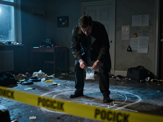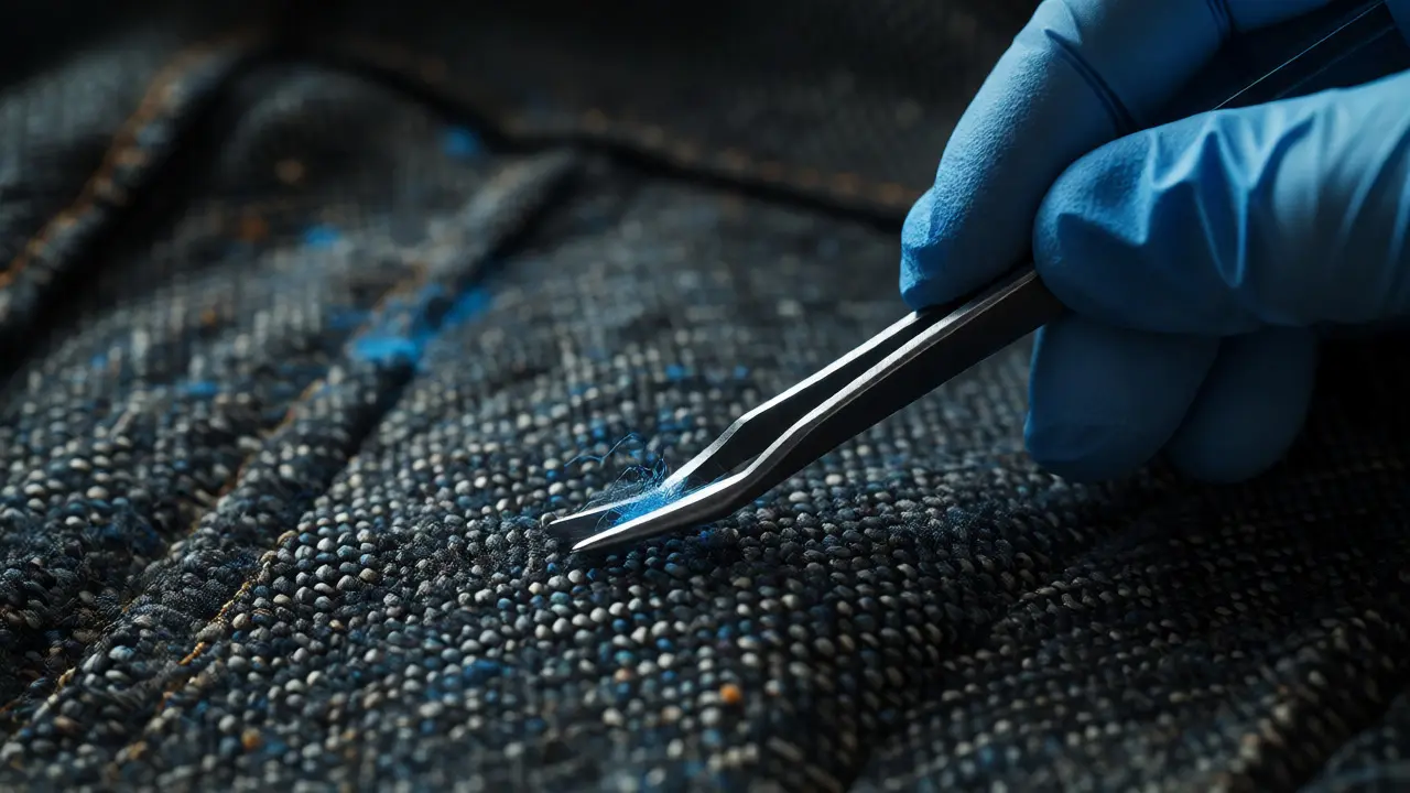
You find a speck of blue fiber on a suspect's jacket. It’s smaller than a grain of sand. To the naked eye, it’s nothing. To a forensic scientist, it’s a potential smoking gun. But here is the catch: if you touch it with dirty tweezers or blow on it while examining it, that "smoking gun" becomes worthless dust. Microscopic evidence is fragile. It requires a specific mindset and a rigorous protocol to survive the journey from the crime scene to the lab bench.
Handling small particles isn’t just about looking closely; it’s about preserving context. A single pollen grain can place a suspect at a specific location. A fragment of paint can link a hit-and-run driver to a victim’s car. But these links only hold up in court if the handling process was flawless. Let’s break down exactly how to handle these tiny clues without destroying them.
The Fragility Factor: Why Small Matters
When we talk about trace evidence, we are usually dealing with materials that weigh less than a milligram. Think hair, fibers, glass shards, gunshot residue (GSR), and soil. The problem isn't just their size; it's their susceptibility to contamination and loss.
Consider glass fragments. If you sweep a window pane with a standard broom, you might collect the glass, but you’ve also mixed it with floor dirt, carpet fibers, and last week’s crumbs. You’ve created a soup. In forensics, separation is everything. You need to isolate the specific particle of interest. Using static charge, specialized vacuums, or careful hand-picking under magnification is often necessary. If you lose the particle, you lose the case. If you contaminate it, you create reasonable doubt.
Collection: The First Line of Defense
Most mistakes happen before the evidence even hits the microscope slide. Collection methods depend entirely on the substrate-the surface where the particle lives. You can’t use the same technique for a wool coat as you do for a smooth vinyl seat.
- Tape Lifting: This is the go-to for dry surfaces like clothing or upholstery. Use low-tack tape to gently lift fibers. High-tack tape can pull the actual material off the fabric, altering its structure.
- Static Charge: For hard, non-porous surfaces like dashboards or floors, a charged balloon or specialized electrostatic device can attract light particles like hair and pollen without touching them.
- Vacuuming: Specialized forensic vacuums use filters to trap particles. Never use a household vacuum. The turbulence inside a home unit destroys delicate structures and mixes samples from different rooms.
- Hand Picking: Under a stereomicroscope, using forceps with blunt tips, analysts can pick out visible particles. This is slow but precise. It’s used when bulk collection would dilute the sample too much.
A critical rule: always wear gloves. Not just any gloves, but lint-free nitrile gloves. Latex gloves shed powder. Cotton gloves shed fibers. If you drop a cotton fiber onto your evidence bag, and the suspect’s shirt is cotton, good luck proving which one came from where.
Packaging: Stop Using Envelopes for Everything
This is where many junior technicians stumble. We love paper envelopes because they breathe. But put a microscopic glass shard in a paper envelope, shake it around, and what happens? The paper abrasion creates new fibers. The shaking grinds the glass into dust. You’ve altered the evidence.
| Evidence Type | Best Container | Why? |
|---|---|---|
| Hair & Fibers | Paper Bindle or Glassine Paper | Prevents static cling; allows visibility without opening. |
| Glass & Paint Chips | Rigid Plastic Vial or Cardboard Box | Protects against crushing; prevents abrasion. |
| Soil & Dust | Sealed Plastic Bag or Jar | Prevents drying out (if wet) or spilling (if dry). |
| Gunshot Residue | Sticky Stub Kit or Swab Tube | Maintains chemical integrity for SEM-EDX analysis. |
For extremely small items, like a single seed or insect part, use a "pill box" style container lined with clean foam or tissue. Secure it with tape so it doesn’t roll around. Label everything immediately. "Blue fiber" is not a label. "Blue fiber collected from left shoulder of suspect’s jacket, Sept 20, 2026, by Officer Smith" is a label.
Lab Analysis: Choosing the Right Microscope
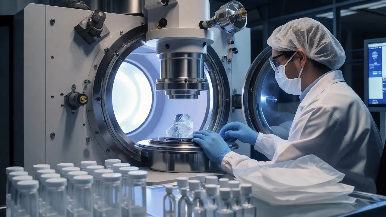
Once the evidence reaches the lab, the choice of instrument determines what you can learn. There is no single "best" microscope. It depends on the question you’re asking.
Comparison Microscopes are dual-headed instruments that let you view two samples side-by-side through a single eyepiece. This is the gold standard for matching bullets, tool marks, or hair morphology. You adjust the focus and position until the features align perfectly. It’s subjective, yes, but highly effective for physical matching.
For chemical composition, you need more than optics. You need Scanning Electron Microscopy (SEM) coupled with Energy Dispersive X-ray Spectroscopy (EDS). An SEM uses electrons instead of light, providing massive depth of field and high magnification (up to 100,000x). When combined with EDS, it tells you the elemental makeup. Is that green fleck copper arsenite pigment? Or is it just oxidized iron? The SEM-EDX combo answers that instantly.
Don’t forget Polarized Light Microscopy (PLM). This is crucial for fibers and minerals. By rotating polarizing filters, you can determine if a fiber is synthetic (like nylon) or natural (like cotton) based on how it interacts with light. PLM is non-destructive, meaning you can keep the sample for DNA testing later if needed.
Contamination Control: The Invisible Enemy
Contamination is the silent killer of forensic credibility. It happens when foreign material gets introduced to the evidence. Sources include the analyst’s breath, skin flakes, airborne dust, or previous samples on the microscope stage.
To combat this, labs follow strict protocols:
- Clean Bench Work: Wipe down all surfaces with 70% ethanol or bleach between cases. Use disposable pipette tips and spatulas.
- Blank Samples: Always run a "blank"-a sample known to be free of target analytes-alongside the evidence. If your blank shows blue fibers, your equipment is contaminated.
- Separate Areas: Pre-analysis (collection/packaging) should never happen in the same room as post-analysis (chemical testing). Airflow should move from clean to dirty areas, not vice versa.
- Personal Hygiene: No talking over open samples. Hairnets are mandatory. Even a single eyelash falling into a vial can confuse results.
Remember, the burden of proof includes proving that the evidence wasn’t contaminated during handling. If you can’t demonstrate a clean workflow, defense attorneys will tear your findings apart.
Documentation and Chain of Custody
Every time a particle moves, it must be logged. From the moment it’s lifted off a jacket to the moment it’s photographed under an SEM, there must be an unbroken chain of custody. This isn’t just paperwork; it’s legal armor.
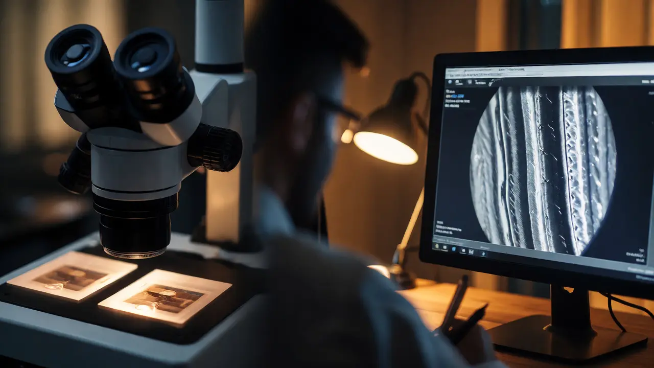
Digital photography has revolutionized this step. Modern digital microscopes capture images directly to the computer. These images become part of the permanent record. They show the scale bar, the lighting conditions, and the exact orientation of the sample. If a dispute arises years later, those raw image files can be re-analyzed. Without them, you’re relying on memory or sketches, which are far less persuasive.
Ensure your notes answer five questions for every action: 1. Who touched it? 2. When did they touch it? 3. Where was it stored? 4. What was done to it? 5. Why was it done? If you miss one, the evidence is compromised.
Common Pitfalls to Avoid
Even experienced professionals make errors. Here are the big ones:
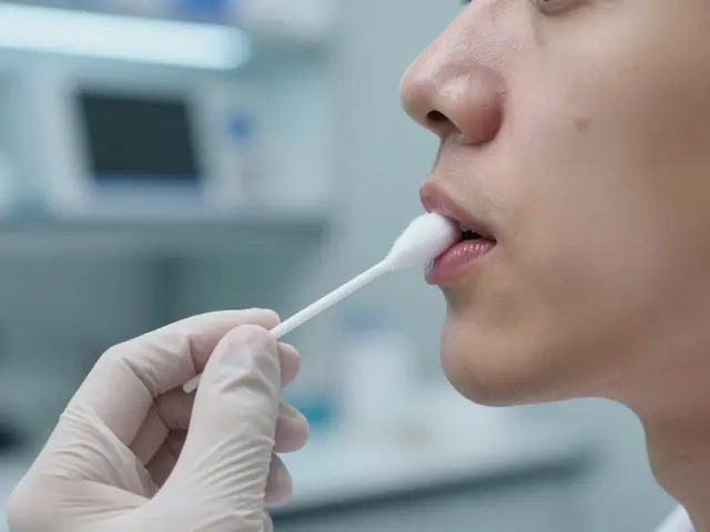
- Over-cleaning: Scrubbing a suspect’s hands to remove GSR before swabbing can wash away the evidence. Gentle swabbing is key.
- Using the wrong solvent: Acetone dissolves many plastics. If you clean a plastic fragment with acetone, you might dissolve the very polymer you’re trying to identify.
- Losing the substrate: Sometimes the value isn’t just in the particle, but in how it’s attached. Pulling a fiber off a cloth loses information about weave direction and tension. Photograph before removing.
- Ignoring background: Collect control samples. If you find pollen on a shoe, you need pollen from the suspect’s house, the victim’s house, and the crime scene. Without controls, you can’t say the pollen is unique to the scene.
Handling microscopic evidence is a discipline of patience and precision. It’s tedious work, often involving hours of staring into lenses. But when that tiny blue fiber matches the victim’s sweater under the comparison microscope, the tedium pays off. It turns a hunch into fact. Treat every speck like it’s the most important thing in the world, because in that moment, it is.
Can microscopic evidence prove guilt beyond a reasonable doubt?
Rarely on its own. Microscopic evidence is typically class evidence, meaning it can link a suspect to a scene or object but cannot uniquely identify an individual like DNA does. However, when combined with other evidence (like video or witness testimony), it strengthens the overall case significantly. A match in fiber type, color, and chemical composition makes coincidence less likely, but it usually supports a narrative rather than standing alone as definitive proof.
How long can microscopic evidence be stored?
Indefinitely, if stored correctly. Dry organic materials like hair and fibers can last decades if kept dark, dry, and cool. Chemical degradation is the main enemy. UV light breaks down dyes in fibers, and humidity causes mold or rust. Proper archival packaging-acid-free paper, sealed containers, controlled environments-is essential for long-term preservation.
What is the difference between trace evidence and microscopic evidence?
They overlap heavily. Trace evidence refers to the *quantity* and *origin* of the material (small amounts transferred during contact). Microscopic evidence refers to the *method* of analysis (requiring magnification). All microscopic evidence is not necessarily trace (e.g., a large gemstone examined under a loupe), and not all trace evidence is microscopic (e.g., a large blood stain). However, in practice, most trace evidence is analyzed microscopically.
Does washing clothes destroy microscopic evidence?
Yes, generally. Washing removes loose fibers, pollen, and soil. Agitation in a washing machine breaks fibers and redistributes particles. Drying adds heat and friction, further degrading delicate materials. While some deeply embedded particles might survive, the evidentiary value drops dramatically after laundering. That’s why collecting unworn clothing from suspects is prioritized.
Is DNA analysis possible on microscopic particles?
Sometimes. Touch DNA can be recovered from items handled by individuals, even if no visible biological material is present. However, success rates vary wildly. Skin cells shed naturally, but environmental factors degrade DNA quickly. For microscopic particles like hair shafts, mitochondrial DNA is often used because nuclear DNA is scarce in the shaft itself. It’s challenging but increasingly common in modern labs.