When a crime scene yields a single hair, a speck of skin, or a cluster of cells too small to see with the naked eye, traditional methods often fail. That’s where laser microdissection steps in - turning invisible clues into usable evidence. This isn’t science fiction. It’s a real, widely used technique in forensic labs today, letting researchers pull out individual cells from complex tissue samples with surgical precision. No hands. No contamination. Just a laser, a microscope, and a target.
How Laser Microdissection Works
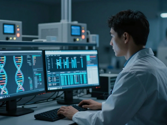
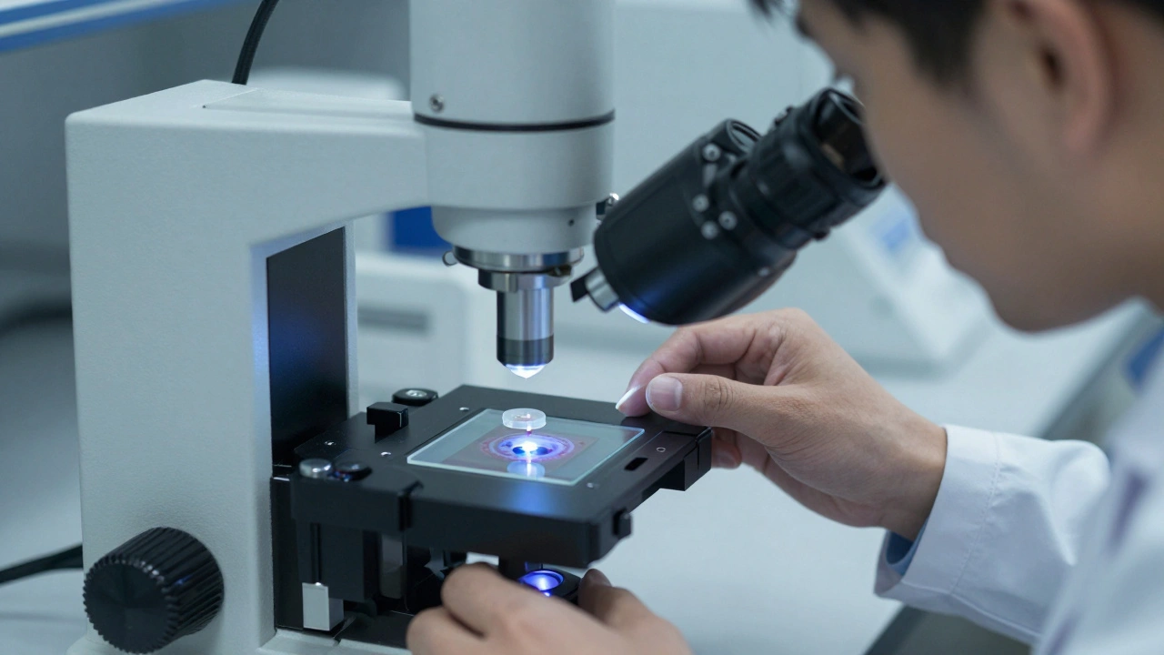
Laser microdissection (LMD), sometimes called laser capture microdissection, uses a focused laser beam to cut and lift out specific cells from a tissue slide. Imagine looking through a microscope at a piece of a tumor, a bloodstain, or a scrap of skin under a fingernail. You see a mix of cell types - some from the victim, some from the suspect, maybe even environmental debris. You want just one kind. LMD lets you pick it out. The process starts with a tissue sample mounted on a special slide. These aren’t regular glass slides. They’re coated with a thin, transparent membrane that holds the sample in place while letting laser light pass through. Under the microscope, the scientist identifies the exact cells they need - maybe 10 cancer cells in a sea of healthy ones, or a single epithelial cell from a bite mark. Then, using a mouse or touchscreen, they draw a shape around those cells. The laser fires. There are two main laser types used. One is ultraviolet (UV), which cuts like a scalpel. It vaporizes tissue along the drawn line, cleanly separating the target. The other is infrared (IR), which works differently. Instead of cutting, it heats a thin thermoplastic film on a cap placed over the sample. The heat melts the film just enough to stick to the selected cells. When the cap lifts, the cells come with it. Both methods avoid touching the sample, which keeps everything pristine. Precision matters. Systems like the MMI CellCut can cut with accuracy down to 0.3 micrometers. That’s less than the width of a human hair. At that scale, you’re isolating individual cells - even chromosomes - without disturbing neighbors. The whole process, from slide prep to collection, usually takes 2 to 8 hours depending on how many cells you need and how complex the tissue is.Why It Matters for Trace Evidence
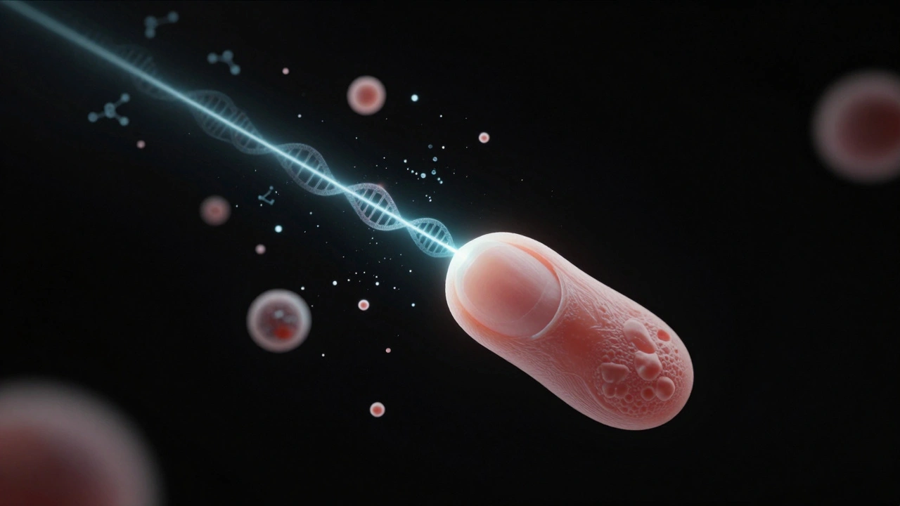
In forensics, trace evidence is everything. A drop of sweat on a doorknob. A strand of hair caught in a zipper. A fleck of skin under a victim’s fingernail. These aren’t just clues - they’re potential DNA sources. But here’s the problem: most samples are messy. A single cell cluster might contain 50 different cell types. If you analyze them all together, you get a jumbled genetic profile. You might miss the suspect’s DNA entirely. Laser microdissection solves that. It lets you isolate only the cells you care about. For example, in a sexual assault case, a swab might contain vaginal epithelial cells, sperm cells, and skin cells from the suspect. Without LMD, labs get a mix. With it, they can isolate just the sperm cells - the ones that likely came from the perpetrator - and run a clean DNA test. No background noise. No false leads. It’s not just about DNA. LMD preserves RNA and proteins too. That means you can study gene expression in tumor cells from a murder victim’s skin, or detect toxins in a single cell from a suspected poisoning case. You’re not just identifying who was there - you’re learning what they were doing, what they were exposed to, even what drugs they’d taken.Key Systems in Use Today
Not all LMD systems are the same. Two major designs dominate forensic and research labs. Leica Microsystems uses a UV laser mounted on an upright microscope. The laser moves to cut tissue, and the dissected material falls into a collection tube by gravity. It’s reliable, but it only works well within the microscope’s field of view. If your target cells are outside that area, you have to reposition the slide - which adds time and risk of error. Molecular Machines International (MMI) flipped the design. Their CellCut system keeps the laser fixed and moves the stage instead. This means the laser stays perfectly in focus no matter where you are on the slide. It’s faster, more accurate, and better for large or scattered samples. They also use a patented adhesive cap that seals over the tissue. This stops tiny cells from floating away due to air currents or static - a common issue with dry samples. Their software, CellTools, even helps identify rare cells automatically using image analysis. That’s huge when you’re looking for one cell in a thousand. Carl Zeiss and Arcturus systems use similar IR laser approaches, melting adhesive films to capture cells. Some even use special slides coated with energy-transfer layers that literally shoot cells into collection tubes when the laser hits. These are contact-free methods - no physical contact means no contamination.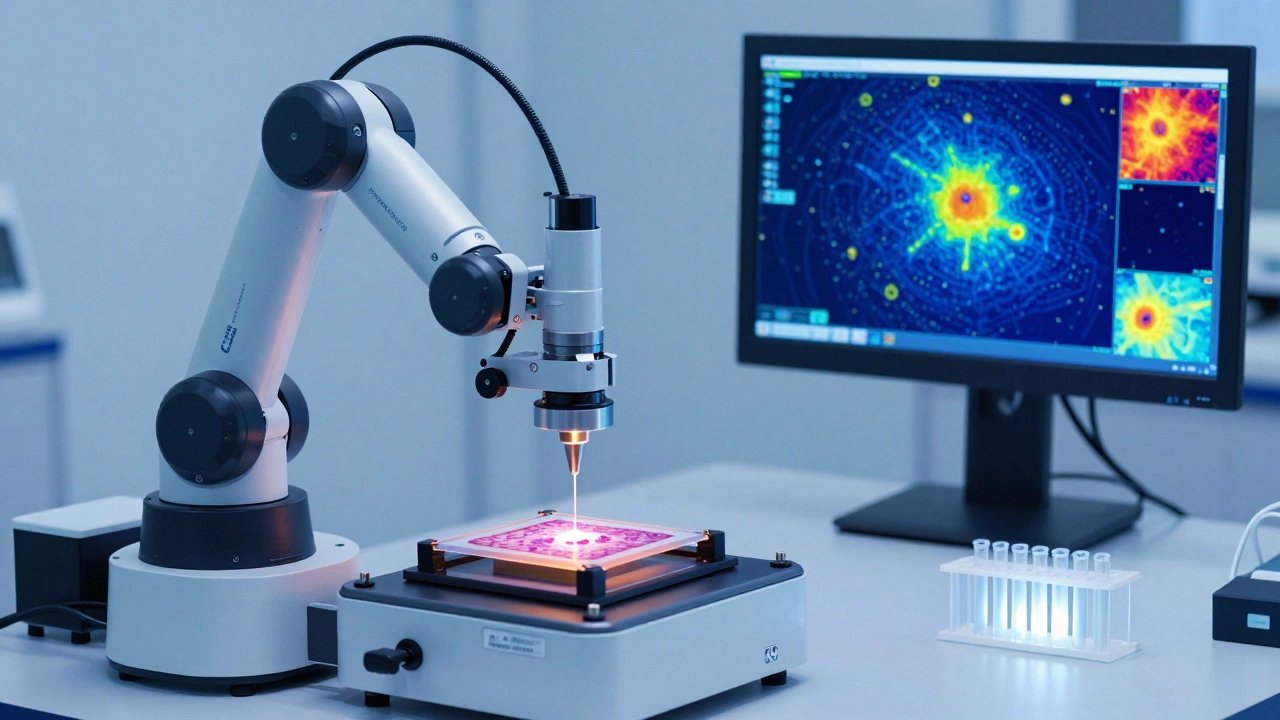
What You Can Do With Isolated Cells
Once you’ve isolated your microscopic target, the real work begins. The collected cells go into standard lab processes:- Genomics: Extract and sequence DNA to match suspects or identify unknown victims.
- Transcriptomics: Analyze RNA to see which genes were active - useful in determining if a cell came from a tumor, an infection, or a drug reaction.
- Proteomics: Identify proteins present, which can reveal age, sex, or even the type of tissue (e.g., lung vs. skin).
- Lipidomics and Metabolomics: Detect fatty acids or metabolic byproducts that might point to specific substances, like explosives or synthetic drugs.
Advantages Over Traditional Methods
Before LMD, forensic labs relied on macro-dissection - cutting tissue with scalpels under a microscope. It was messy. You’d drag cells, contaminate samples, or lose material. Even micro-pipetting couldn’t isolate single cells reliably. LMD changes that:- Pure samples: Only the cells you select are collected. No cross-contamination.
- Preserved integrity: DNA, RNA, and proteins stay intact. No degradation from physical handling.
- High precision: Isolate single cells or even parts of chromosomes.
- Non-contact: No tools touch the sample. No risk of introducing foreign material.
- Scalable: You can pick 1 cell or 1,000 in one session.
Limitations and Challenges
LMD isn’t perfect. It’s expensive. A single system costs over $150,000. Not every lab can afford one. Training takes months. You need skilled operators who understand both microscopy and molecular biology. Sample prep is tricky. Tissue must be thin enough to cut - usually 5-10 micrometers thick. Too thick, and the laser can’t reach. Too thin, and you lose structural context. Fixation matters too. Over-fixing with formalin can destroy RNA. Under-fixing leaves cells disintegrating. And there’s still human error. If the operator misidentifies a cell - maybe confusing a fat cell for a cancer cell - the whole analysis is off. That’s why newer systems are adding AI-assisted cell detection. They scan the slide, highlight patterns, and suggest targets. Human eyes still confirm, but the computer does the heavy lifting.The Future of LMD in Forensics
The next five years will bring big changes. Automation is coming. Labs are testing systems that can scan entire slides, auto-select target cells, and run pre-programmed dissections without human input. That could cut analysis time from hours to minutes. Integration with portable DNA sequencers is another frontier. Imagine a crime scene unit that uses LMD to isolate a single cell, then runs a full genome sequence on-site in under an hour. No need to send samples to a central lab. Faster results. Faster justice. And as costs drop, LMD will move beyond elite labs. Universities, smaller forensic centers, and even international agencies are starting to adopt it. What was once a niche tool is becoming standard.What This Means for Justice
In a world where evidence is often ambiguous, LMD brings clarity. It turns vague traces into concrete links. A single cell can convict. Or exonerate. It doesn’t replace fingerprints or surveillance. But it adds a layer of molecular precision that no other technique can match. When every micrometer counts, laser microdissection isn’t just useful - it’s essential.Can laser microdissection be used on old evidence?
Yes, but with limitations. LMD works best on samples preserved with proper fixation - usually formalin or frozen tissue. Older samples that were dried out, exposed to heat, or improperly stored may have degraded DNA or RNA, making analysis harder. However, even degraded samples can sometimes yield usable genetic material if the target cells are still structurally intact. Labs often test multiple areas of old slides to find the best-preserved regions.
How long does it take to get results from laser microdissection?
The actual dissection takes 2 to 8 hours, depending on sample complexity. But the full timeline includes sample prep (1-2 days), dissection (several hours), and downstream analysis like DNA sequencing (another 1-5 days). So total turnaround is typically 3 to 7 days. Fast-track labs using automation can reduce this to under 48 hours for urgent cases.
Is laser microdissection used in criminal trials?
Yes. Courts in the U.S., Europe, and Australia have accepted LMD-derived DNA evidence in over 200 criminal cases since 2018. The technique is validated under ASTM and ISO standards for forensic DNA analysis. Its repeatability, precision, and contamination control make it legally defensible. Expert testimony often explains how the laser isolates only the target cells, eliminating doubt about sample purity.
Can LMD identify the sex or age of a person from a single cell?
Yes. From a single cell, labs can extract DNA and look for sex chromosomes (XX or XY) to determine biological sex. Age estimation is trickier but possible through epigenetic markers - chemical changes on DNA that accumulate over time. Some labs now use LMD to isolate skin cells from bite marks and estimate the donor’s age within a 5-10 year range, which helps narrow suspect pools.
What’s the smallest sample LMD can handle?
The smallest samples are single cells - sometimes even fragments of a cell. LMD systems can isolate cells as small as 5 micrometers in diameter. That’s about one-tenth the width of a human hair. In forensic cases, this means even a single epithelial cell from a touch DNA sample can be collected and analyzed, which was impossible just 15 years ago.