When a person dies under suspicious circumstances, the body becomes the primary witness. It holds the physical proof of what happened, but that proof is fragile. If you don't handle it correctly, critical evidence disappears forever. This is where a forensic autopsy comes in. Unlike a standard hospital autopsy, which looks for medical causes, this procedure is designed to answer legal questions. It determines how and why someone died while preserving every scrap of evidence for court.
Getting this right isn't just about medical knowledge; it's about strict adherence to protocol. Organizations like the National Association of Medical Examiners (NAME) have updated their standards as recently as 2025 to ensure these exams are reproducible and defensible. Whether you're a student, a trainee, or just curious about the process, understanding the workflow reveals why precision matters more than speed.
Why Forensic Autopsies Are Different
You might wonder why we can't just use a regular clinical autopsy. The difference lies in the goal. A clinical autopsy helps doctors understand disease progression or treatment errors. A forensic autopsy serves the justice system. Its main jobs are determining the cause of death (the injury or disease that killed the person) and the manner of death (homicide, suicide, accident, natural, or undetermined).
This distinction changes everything. In a forensic setting, every cut, swab, and photograph must follow a chain of custody. You aren't just looking for a heart attack; you're looking for trace fibers on clothing, bite marks on skin, or drugs in the blood that could exonerate or convict someone. Because of this high stakes environment, only board-certified forensic pathologists or supervised trainees should perform these procedures. Letting general pathologists handle them without guidance is considered risky by professional bodies because they may miss subtle signs of trauma or mishandle evidence.
Pre-Autopsy: Context Is King
Before a single incision is made, the work begins with information. According to NAME’s 2025 standards, the pathologist must review the circumstances of death before touching the body. This includes police reports, scene photos, and witness statements. Why? Because knowing that a victim was found in a locked room changes how you look for injuries compared to finding them at a car crash site.
Identification is the first concrete step. For known individuals, this means confirming identity through records and photos. For unidentified bodies, the process is much heavier. The pathologist must take full-body radiographs (X-rays), chart dental records via X-ray, collect fingerprints if possible, and archive DNA samples. These steps happen before the body is altered, ensuring that even if decomposition sets in later, identification data remains intact.
Reception and Initial Documentation
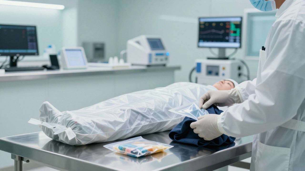
When the body arrives at the morgue, the clock starts ticking on preservation. The very first action is to photograph the decedent "as presented." This means capturing the body exactly as it was received-clothing intact, position unchanged, any coverings in place. This visual record proves that no alterations were made before the examination began.
Next comes the removal of clothing. This isn't just undressing; it's an evidence hunt. Technicians carefully cut away garments to avoid destroying fibers or debris stuck to seams. They measure and record the body’s length and weight immediately after. These anthropometric measurements help identify unknowns and assess nutritional status or drug effects. Any trace evidence found during this phase-like glass shards or soil-is bagged and labeled separately.
The External Examination: Surface Survey
Now the naked body is examined from head to toe. This external survey is systematic. The pathologist documents scars, tattoos, birthmarks, and amputations. These identifying features are crucial for public releases if the body is unclaimed.
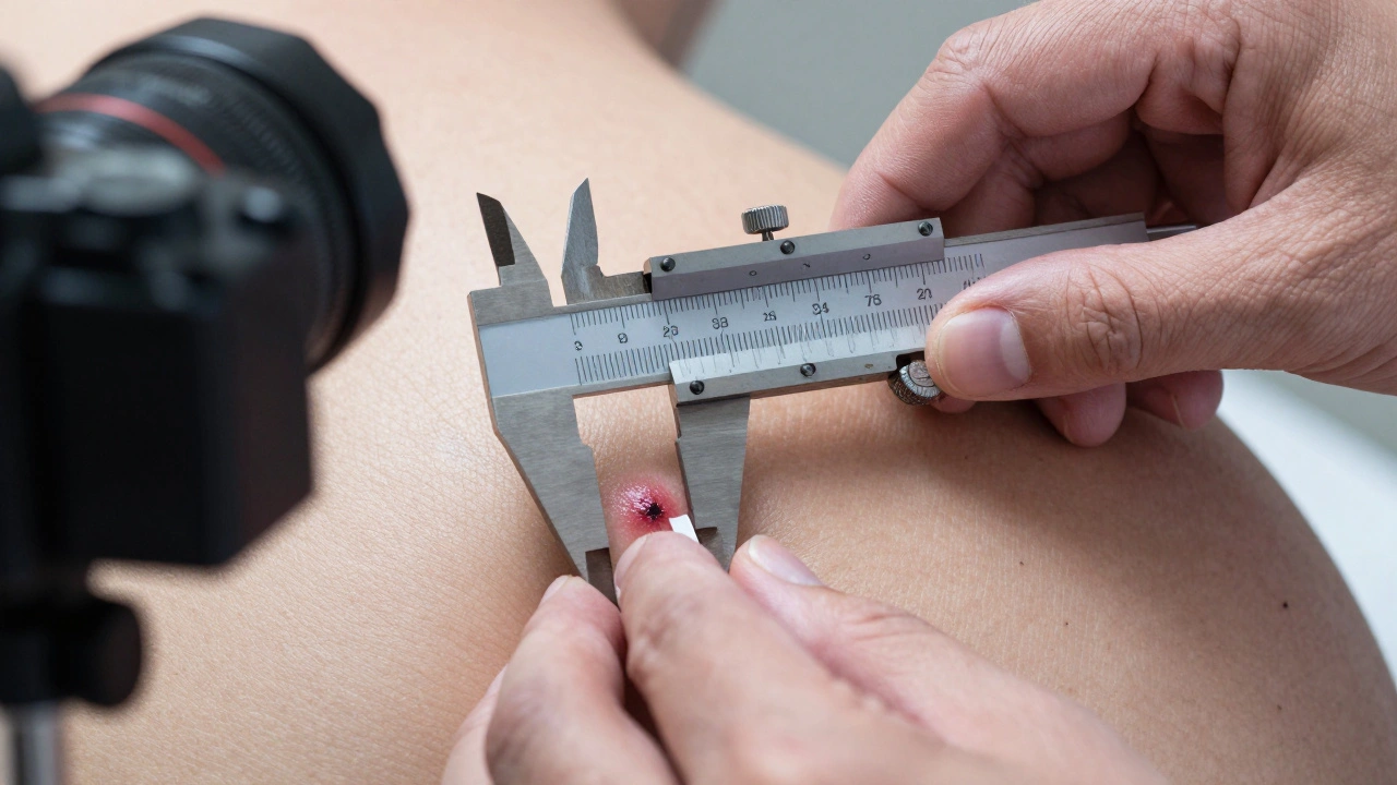
More importantly, they look for signs of life and death processes. Rigor mortis (stiffening) and livor mortis (pooling of blood) help estimate time since death. Decomposition levels tell you how long the body has been exposed. But the focus is on trauma. Every wound is measured using Vernier calipers for millimeter accuracy. Descriptions include type (incised, blunt force, gunshot), location, size, shape, and color. Photographs are taken with scales next to each injury to show true size. If there’s a bite mark, a swab is taken for DNA before cleaning the area. This stage never skips the posterior side of the body; back injuries are common in defensive struggles or restraints.
Collecting Trace and Sexual Assault Evidence
Before washing the body, specific biological evidence must be collected. This is non-negotiable in cases involving potential sexual assault or strangulation. Swabs are taken from the oral, vaginal, and rectal cavities. Pubic hair combings or tape lifts are performed to catch transferred hairs or fibers. Fingernail clippings and scrapings are collected because victims often scratch attackers, leaving skin cells under the nails.
If you wash the body first, you destroy this evidence. That’s why protocols mandate collecting these samples prior to any cleaning. Each sample is packaged in sterile containers, labeled with the anatomical source, and placed into the chain of custody. This ensures that if DNA matches a suspect, the defense cannot claim contamination occurred during the autopsy.
Internal Examination: Dissection and Organ Analysis
With external documentation complete, the internal exam begins. The pathologist makes a Y-shaped incision from shoulder to shoulder down to the pubic bone. Ribs are cut away to access the chest cavity. Organs are removed either individually or en bloc (as a block), depending on the case needs and regional practice.
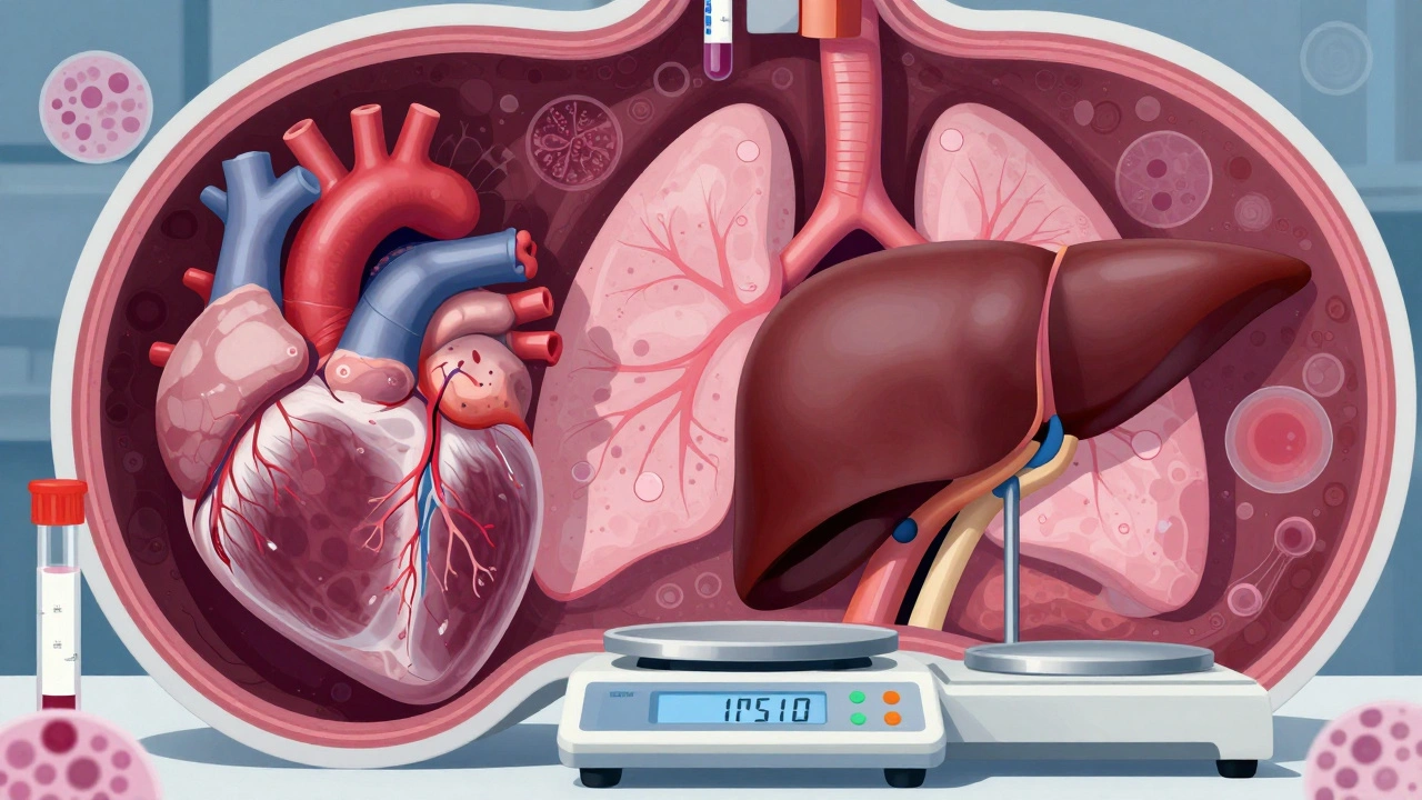
Each organ-the heart, lungs, liver, kidneys, brain-is weighed and inspected grossly. Weights matter. An enlarged heart suggests chronic hypertension; heavy lungs might indicate drowning or pneumonia. The pathologist slices through tissues to check for hidden hemorrhages, tumors, or foreign objects like bullets. Tissue samples are taken for histology (microscopic analysis). This is vital when the cause of death isn't obvious, such as in sudden infant death syndrome or suspected drug overdoses where tissue inflammation provides clues.
Ancillary Studies: Radiology, Toxicology, and Histology
Autopsy doesn't end with the knife. Three key lab tests support the findings:
- Radiology: X-rays or CT scans are used, especially in unidentified cases or trauma, to find fractures, projectiles, or implants that aren't visible externally.
- Toxicology: Blood, urine, and vitreous fluid (from the eye) are collected. Vitreous fluid is particularly stable and useful for detecting glucose, electrolytes, and toxins even in decomposed bodies. Samples are sent to a lab to test for alcohol, prescription drugs, illicit substances, and poisons.
- Histology: Microscopic slides of tissues reveal cellular changes. For example, soot in lung tissue confirms inhalation of smoke in fire deaths, distinguishing live burns from postmortem burning.
Documentation and Reporting
The final product is the autopsy report. This isn't just a summary; it's a legal document. NAME standards require a narrative report including the date, time, location, and names of all personnel involved. It must detail external and internal findings, list diagnoses, and explicitly state the cause and manner of death. The pathologist signs and dates it, taking personal responsibility for the conclusions.
Accurate reporting prevents misinterpretation in court. Vague terms like "possible" or "likely" are avoided unless justified. Instead, precise language links findings to mechanisms of death. For instance, instead of saying "heart failure," a report might specify "acute myocardial infarction due to coronary artery thrombosis." This clarity allows prosecutors and defense attorneys to build their cases on solid facts.
| Feature | Clinical Autopsy | Forensic Autopsy |
|---|---|---|
| Purpose | Medical education, quality improvement | Determine cause/manner of death, legal evidence |
| Authority | Next-of-kin consent | Coroner/Medical Examiner order |
| Evidence Handling | Minimal, not required | Strict chain of custody, trace evidence collection |
| Performed By | Anatomical pathologist | Forensic pathologist |
| Report Focus | Disease processes | Trauma, toxicology, legal conclusions |
Challenges and Best Practices
One major challenge is decomposition. When bodies are severely degraded, traditional methods fail. In these cases, radiology and toxicology become even more critical. Another issue is bias. Pathologists must remain objective, avoiding assumptions based on media reports or police theories. The 2025 NAME standards emphasize reviewing scene data to contextualize findings but warn against letting preconceptions dictate results.
Training is essential. New forensic pathologists undergo years of residency and fellowship. They learn to recognize subtle signs of abuse, differentiate antemortem from postmortem injuries, and interpret complex toxicology results. Continuous updates to guidelines ensure practices evolve with new technologies, like virtual autopsy (virtopsy) using CT/MRI, which is gaining traction as a complementary tool.
Who performs a forensic autopsy?
A board-certified forensic pathologist performs the autopsy. In some jurisdictions, trained residents or fellows may assist under direct supervision. General pathologists without forensic training should not conduct these exams independently due to the specialized nature of evidence handling and trauma interpretation.
How long does a forensic autopsy take?
A typical forensic autopsy takes 2 to 4 hours. Complex cases involving extensive trauma, decomposition, or additional imaging/toxicology sampling can take longer. The timeline depends on the condition of the body and the number of ancillary tests required.
What is the difference between cause and manner of death?
Cause of death is the specific injury or disease that led to death (e.g., gunshot wound to the head). Manner of death is the classification of the circumstances (e.g., homicide, suicide, accident, natural, or undetermined). Both are determined by the forensic pathologist based on autopsy findings and scene investigation.
Can family members watch the autopsy?
Generally, no. Forensic autopsies are legal proceedings, and privacy/security concerns restrict observation. However, some jurisdictions allow families to view the body before the procedure begins or provide detailed reports afterward. Policies vary by region and agency.
Why are X-rays taken during a forensic autopsy?
X-rays help identify orthopedic implants, dental work, bullets, shrapnel, or fractures that aren't visible externally. They are crucial for identifying unknown decedents and documenting occult injuries that could indicate foul play or accidental trauma.