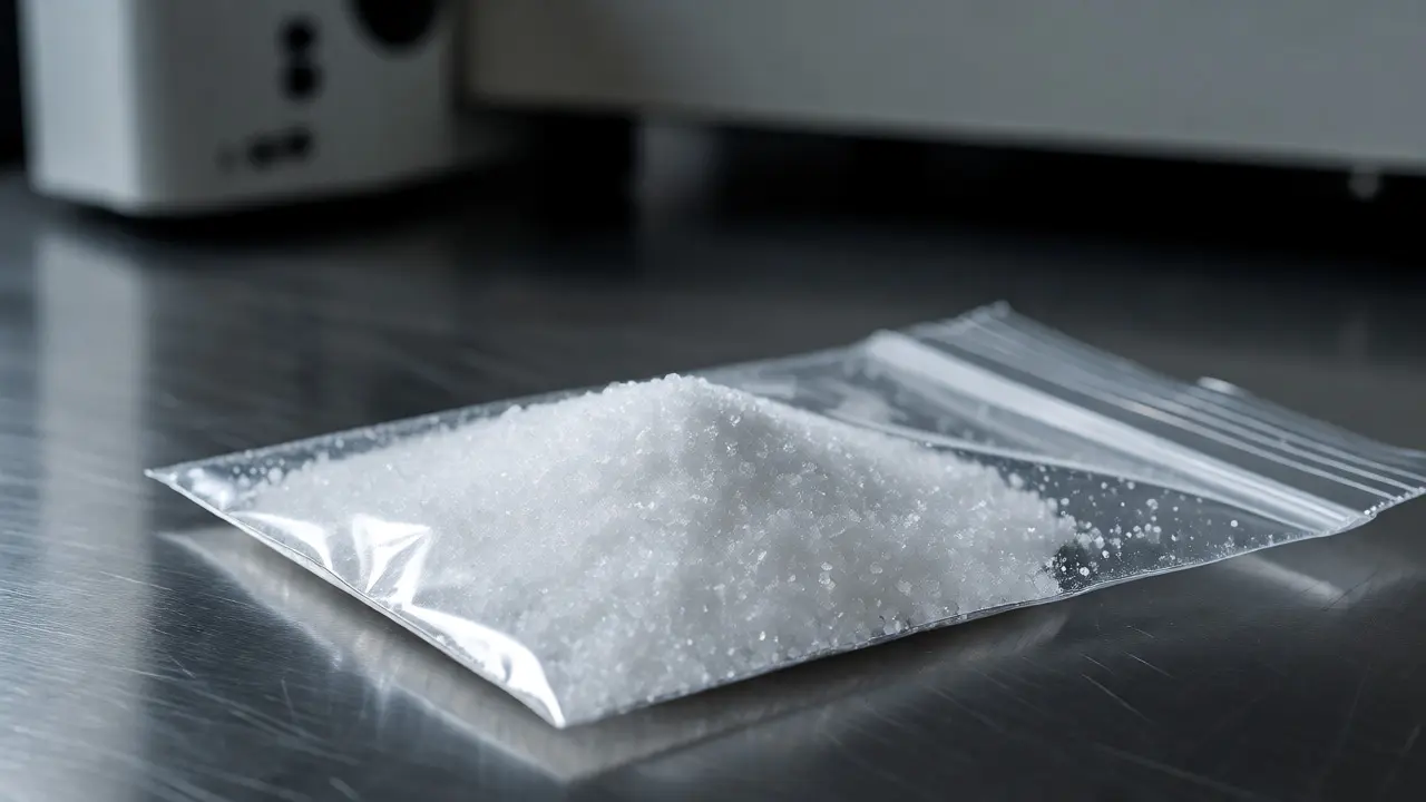
You find a white powder in a sealed evidence bag. Visually, it looks like sugar, salt, or maybe crushed aspirin. But in forensic labs and pharmaceutical quality control, "looks like" is dangerous. Two compounds can have identical chemical formulas yet behave completely differently in the body because their molecules pack together in different crystal shapes. This phenomenon, known as polymorphism, is why one version of a drug might dissolve quickly while another passes through you unchanged.
To tell these versions apart without destroying the sample, analysts turn to two powerful tools: Fourier Transform Infrared Spectroscopy (FTIR) and Raman Spectroscopy. Both probe molecular vibrations, but they do so in ways that complement each other perfectly. If you are trying to identify a controlled substance, verify a pharmaceutical batch, or distinguish between counterfeit pills, understanding how these techniques reveal crystal structure is non-negotiable.
Why Crystal Structure Matters More Than Chemical Formula
Most people think chemistry is just about atoms bonded together. But in solid-state chemistry, how those molecules arrange themselves in space defines the material's identity. Think of ice, water, and steam. Same H₂O formula, totally different properties. Now scale that down to the molecular level within a solid crystal.
Ritonavir is the classic cautionary tale here. In the late 1990s, this HIV drug was sold as a capsule containing Form I crystals. It worked great. Then, during production, a new, more stable form called Form II emerged spontaneously. Form II didn't dissolve properly in the gut. The drug became less effective, causing treatment failures worldwide. Abbott Labs had to recall the product and redesign the formulation.
This isn't rare. Many drugs, including carbamazepine, caffeine, and paracetamol, exist in multiple crystalline forms. Each form has distinct melting points, solubility rates, and stability profiles. For forensic scientists analyzing seized drugs, identifying the specific polymorph can sometimes hint at the manufacturing process or even the geographic origin of the sample. For pharmacists, it determines whether the pill actually delivers the dose promised on the label.
How FTIR Reveals Molecular Fingerprinting
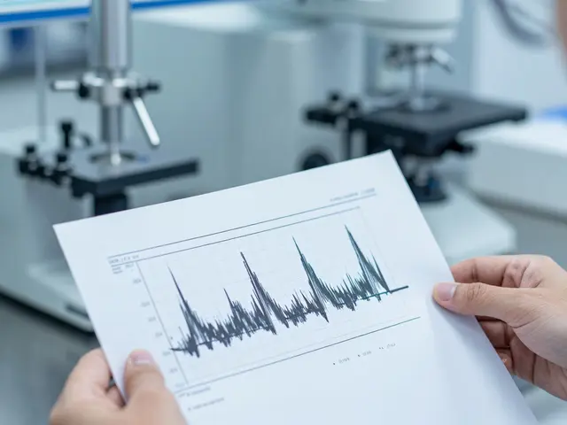
FTIR works by shining infrared light through a sample and measuring which wavelengths get absorbed. Molecules absorb IR energy when the frequency matches their vibrational modes-stretching bonds, bending angles, twisting groups. Because every functional group absorbs at specific frequencies, FTIR creates a unique spectral fingerprint.
In crystal analysis, FTIR shines when distinguishing polymorphs that differ in hydrogen bonding networks. Hydrogen bonds are sensitive to distance and angle. If a drug molecule shifts slightly in its crystal lattice, the O-H or N-H stretching bands shift position or change shape. Analysts look for these subtle changes in the 3200-3600 cm⁻¹ region.
For example, consider Paracetamol (Acetaminophen). Its Form I and Form II show clear differences in the amide I band around 1650 cm⁻¹ due to variations in intermolecular hydrogen bonding. A trained analyst can spot the difference in seconds using standard library matching software, provided the reference spectra include both polymorphs.
However, FTIR has limitations. Water interferes strongly with IR measurements. If your sample is wet or hydrated, the broad O-H stretch from water can mask critical peaks. Also, symmetric vibrations often don't absorb IR well, meaning some structural features remain invisible.
The Complementary Power of Raman Spectroscopy
If FTIR sees asymmetrical vibrations, Raman Spectroscopy excels at detecting symmetrical ones. Raman measures scattered light rather than absorbed light. When laser light hits a sample, most photons bounce off unchanged (Rayleigh scattering), but a tiny fraction exchanges energy with molecular vibrations, shifting their wavelength. This shift reveals the vibrational spectrum.
Raman is particularly useful for drug analysis because water is a weak Raman scatterer. You can analyze aqueous solutions, suspensions, or hydrated crystals without interference. This makes it ideal for checking if a drug has absorbed moisture during storage, which can trigger polymorphic transitions.
Moreover, Raman is highly sensitive to lattice vibrations-collective movements of molecules within the crystal. These low-frequency modes (below 400 cm⁻¹) are often diagnostic for polymorphs but are difficult to detect with standard mid-IR instruments. Modern Fourier Transform Raman (FT-Raman) systems extend into this region, providing a clearer picture of crystal packing efficiency.
Consider Caffeine. Anhydrous caffeine and its monohydrate form look similar under a microscope, but their Raman spectra differ significantly in the ring-breathing modes around 700-800 cm⁻¹. Raman also avoids fluorescence issues better than visible-light Raman when using near-infrared lasers (1064 nm), making it safer for colored or degraded samples.
| Feature | FTIR | Raman |
|---|---|---|
| Interaction Type | Absorption | Inelastic Scattering |
| Water Interference | High (Strong absorber) | Low (Weak scatterer) |
| Best For | Asymmetric stretches, H-bonds | Symmetric stretches, Lattice modes |
| Sample Prep | KBr pellets, ATR, Nujol mull | Minimal (Glass vial, direct contact) |
| Fluorescence Risk | Negligible | Moderate (mitigated by NIR lasers) |

Choosing the Right Technique for Your Sample
Should you run FTIR or Raman? Often, the answer is "both." They follow selection rules that are mutually exclusive for centrosymmetric molecules. If a vibration is active in IR, it is usually inactive in Raman, and vice versa. Combining them gives you the complete vibrational picture.
Start with FTIR if:
- The sample is dry and stable.
- You suspect differences in hydrogen bonding patterns.
- You need rapid screening against a large digital library (NIST SDBS, etc.).
- Budget is a constraint; FTIR instruments are generally cheaper.
Switch to Raman if:
- The sample contains water or is in a glass container.
- You need to detect low-frequency lattice modes.
- The compound is highly fluorescent under visible light (use 1064 nm NIR Raman).
- You need to map spatial distribution of polymorphs across a tablet surface.
In forensic contexts, where chain of custody and non-destructive testing are paramount, portable Raman devices have become game-changers. Officers can scan a suspected methamphetamine package in the field. While field devices may not resolve fine polymorphic details, they confirm the base compound. Back in the lab, high-resolution benchtop FTIR and Raman systems provide the definitive crystallographic data needed for court-admissible reports.
Practical Pitfalls in Polymorph Identification
Even with the best instruments, errors happen. Here are common traps in drug analysis:
1. Phase Transformation During Measurement: Grinding a sample for KBr pellet preparation can induce phase changes. Mechanical stress might convert metastable Form II back to stable Form I. Always use gentle preparation methods like Attenuated Total Reflectance (ATR) for FTIR or direct laser focus for Raman to minimize physical disturbance.
2. Hydrate vs. Anhydrate Confusion: Some drugs form hydrates easily. If you heat a sample too much in an oven-dryer before FTIR, you might strip water away, creating an artificial anhydrous spectrum. Conversely, storing a hygroscopic drug in humid air can create a hydrate that wasn't originally present. Monitor weight loss or gain alongside spectral changes.
3. Library Matching Errors: Standard libraries often contain only the thermodynamically stable form. If you encounter a metastable polymorph, the software might match it poorly or incorrectly assign it to the wrong compound. Always visually inspect key peak positions and intensities. Don't trust the similarity index blindly.
4. Mixtures: Real-world samples are rarely pure. A tablet might contain a blend of polymorphs or be mixed with excipients. Excipients like lactose or microcrystalline cellulose have strong IR/Raman signals that can obscure the API (Active Pharmaceutical Ingredient). Use chemometric tools like Principal Component Analysis (PCA) to separate signal contributions.
Advanced Applications: Mapping and Imaging
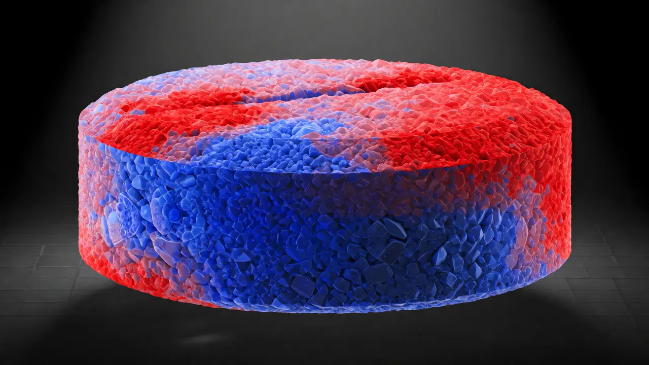
Modern spectrometers don't just give you a single spectrum; they create images. Hyperspectral imaging combines microscopy with spectroscopy. By scanning a grid over a tablet cross-section, you generate a map showing exactly where each polymorph resides.
This is crucial for understanding dissolution behavior. If Form I is concentrated on the surface and Form II is inside, the drug might dissolve initially fast then slow down dramatically. Imaging reveals this stratification. In forensic science, this helps determine if a mixture of powders was physically blended or co-crystallized, offering clues about how a drug was cut or adulterated.
Another emerging trend is coupling these techniques with X-ray Diffraction (XRD). While XRD provides the gold-standard unit cell parameters, it requires larger amounts of pure powder and takes longer. FTIR and Raman serve as rapid triage tools. If they flag a potential polymorphic anomaly, you send the sample for XRD confirmation. This workflow saves time and resources.
Next Steps for Analysts
If you are setting up a protocol for crystal structure analysis in drug cases, start by building a robust local reference library. Do not rely solely on commercial databases. Synthesize or obtain certified reference materials for the specific polymorphs relevant to your caseload. Document the conditions under which each spectrum was collected-temperature, humidity, instrument resolution.
Train your staff to recognize the characteristic signatures of common polymorphs. Teach them to question automated results. And remember: no single technique tells the whole story. Triangulate your findings with melting point analysis, DSC (Differential Scanning Calorimetry), and visual microscopy whenever possible.
Can FTIR and Raman definitively identify all polymorphs?
Not always alone. While they are excellent for distinguishing many polymorphs, some have very similar vibrational spectra. In such cases, X-ray Powder Diffraction (XRPD) is required for definitive proof because it directly probes long-range order and unit cell dimensions. FTIR and Raman are best used as rapid screening tools to flag differences.
Which technique is better for analyzing tablets with coatings?
Raman is generally superior for coated tablets because the laser can penetrate thin polymer coatings (like Opadry) without significant interference. FTIR, especially with ATR accessories, often only samples the surface coating unless you crush the tablet or use transmission mode with careful sectioning.
Does hydration state affect the choice between FTIR and Raman?
Yes. Water strongly absorbs IR radiation, potentially obscuring drug peaks in FTIR. Raman is insensitive to water, making it the preferred method for analyzing hydrated salts or samples stored in humid environments. However, note that the presence of water itself changes the crystal structure, so you must interpret the spectra knowing whether the sample is hydrated or anhydrous.
How does polymorphism impact drug efficacy?
Different polymorphs have different lattice energies and solubilities. A metastable polymorph typically dissolves faster, leading to higher bioavailability, but it may convert to a stable form during storage, reducing efficacy. A stable polymorph dissolves slower but remains consistent over time. Incorrect polymorph selection can lead to therapeutic failure or toxicity.
Are there safety concerns when using lasers in Raman analysis of drugs?
Generally, no, provided standard laser safety protocols are followed. The laser power is usually low (milliwatts). However, some photosensitive drugs may degrade under prolonged laser exposure. It is advisable to minimize dwell time and avoid focusing the laser on the same spot for extended periods if degradation is a concern.