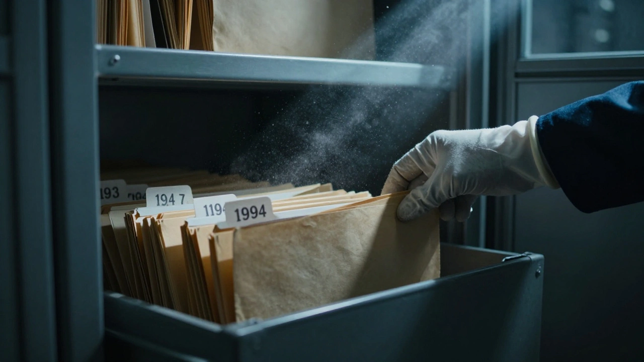
Imagine a file sitting in a dusty cabinet for thirty years. The victim’s name is faded on the tab. The leads are dead ends. The family has moved on, or perhaps they never stopped hoping. For decades, these files were considered closed-not because justice was served, but because there was simply nothing left to do. But today, that narrative is changing. Thanks to breakthroughs in cold case pathology, investigators can now look at old evidence with entirely new eyes.
We aren’t just talking about better microscopes. We’re talking about a fundamental shift in how we understand death, decomposition, and biological traces. A body buried for two decades isn’t just rotting flesh; it’s a time capsule of data waiting for the right key to unlock it. From scanning skeletal remains without digging them up to pulling usable DNA from formalin-soaked tissue blocks, modern forensics is rewriting the rules of what’s possible.
The Scale of the Problem: Why Cold Cases Matter Now
To understand why this matters, you have to look at the numbers. According to the National Institute of Justice (NIJ), there are more than 242,000 unsolved homicides in the United States alone. That’s not a small backlog; it’s a massive reservoir of unresolved trauma for families and communities. Most agencies define a "cold case" as one where all initial leads have been exhausted, often after a specific time threshold like one, three, or five years.
For a long time, once a case went cold, it stayed that way. The original autopsy report was filed, the evidence bagged, and the investigation stalled. But if even 5% to 10% of those 242,000 cases still have retained biological materials-like paraffin blocks, histology slides, or organ samples stored in formalin-we’re looking at tens of thousands of opportunities for re-examination. The goal isn’t just to solve crimes for the sake of statistics; it’s to correct the record. Sometimes, the original cause of death was wrong. Sometimes, the manner of death was misclassified. And sometimes, a tiny piece of overlooked evidence holds the key to identifying a killer who thought they’d gotten away with it.
Seeing the Unseen: Advanced Postmortem Imaging
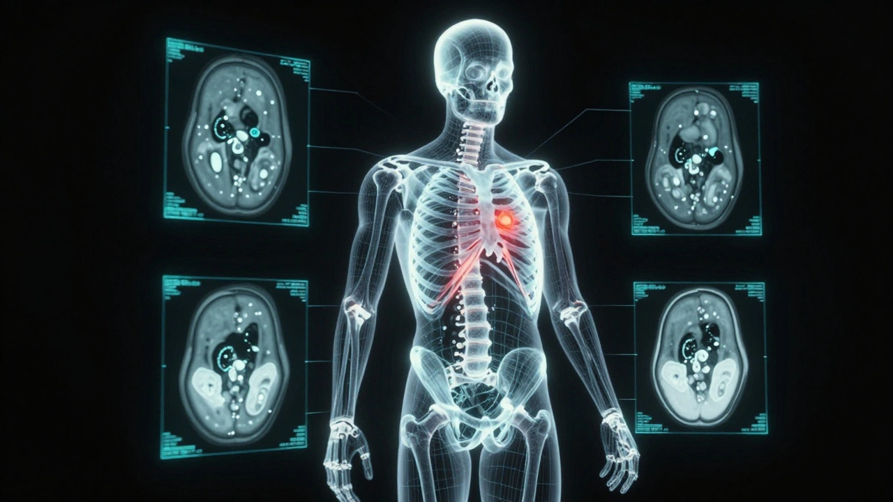
One of the most dramatic shifts in recent years is the adoption of advanced imaging, specifically postmortem computed tomography (PMCT) and magnetic resonance imaging (MRI). In the past, examining a decomposed body or skeletal remains required invasive dissection, which could destroy fragile evidence. Today, teams like the "Virtopsy" group in Switzerland have shown that multislice CT scanners can capture sub-millimeter slices of an entire body in under ten minutes.
This technology allows forensic pathologists to create detailed 3D reconstructions of injuries without ever cutting into the body. Imagine a skeleton buried for twenty years. With PMCT, experts can trace bullet trajectories through bone, identify small knife nicks on ribs, or spot old fractures that were previously mistaken for postmortem damage caused by burial pressure. It’s non-destructive, highly precise, and provides a permanent digital record that multiple experts can review independently. This is crucial for cold cases where the physical evidence is degraded, and every detail counts.
Re-reading the Tissue: Histology and Immunohistochemistry
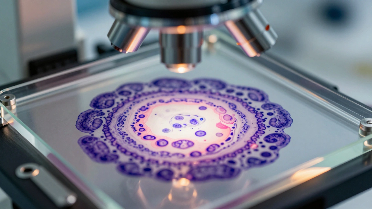
Beyond bones and bullets, soft tissue tells a story too. Many cold cases have archived histology slides-thin slices of tissue stained with dyes and preserved in glass slides for decades. These slides were originally examined using routine stains like hematoxylin and eosin (H&E). But modern immunohistochemistry (IHC) allows pathologists to apply specific markers that reveal details invisible before.
Consider a case involving alleged strangulation. Did the neck injuries happen before death, or were they artifacts of decomposition? By applying markers like CD68 for macrophages or GFAP for astroglial reactions, pathologists can detect vital reactions-signs that the body fought back while alive. If inflammatory cells are present around the hyoid bone or in neck muscles, it suggests antemortem trauma. If they’re absent, it might point to postmortem changes. This distinction is critical. It can turn an ambiguous death into a clear homicide, or conversely, exonerate a suspect by proving injuries occurred after death.
A notable example involves the re-examination of Theresa Ann Green’s case. Forensic pathologist Jonathan Arden reviewed existing autopsy photos and materials and concluded that apparent neck and facial changes were consistent with decomposition rather than hemorrhage from strangulation. He noted that facial bloating often occurs early and prominently, and gravity can pull decomposition fluids down onto the chest, mimicking injury patterns. Without this updated understanding of taphonomy-the study of decay-earlier interpretations might have persisted unchecked.
Toxicology in the Digital Age
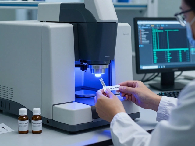
Toxicology has undergone a revolution. In the 1970s or 80s, toxicology screens might have checked for 10 to 20 common drugs using methods with relatively high detection limits. Today, liquid chromatography-tandem mass spectrometry (LC-MS/MS) can detect hundreds of analytes at nanogram-per-milliliter levels. This sensitivity extends to unconventional matrices like hair, nails, and even preserved tissues.
In cold cases, exhumed bodies or archived samples can yield surprising results. Hair segments, for instance, grow at a predictable rate-about one centimeter per month. By analyzing hair in 1-cm intervals, toxicologists can reconstruct a timeline of drug exposure over months or years. This is invaluable for determining if a victim was poisoned chronically or acutely. However, caution is needed. Formalin fixation and long-term storage can degrade certain compounds, so labs must validate their methods carefully. But when done right, modern toxicology can uncover poisons that were completely missed in the original investigation.
DNA from the Past: FFPE Samples and Genetic Genealogy
Perhaps the most powerful tool in the cold case arsenal is DNA analysis. While short tandem repeat (STR) profiling became standard in the 1990s, getting quality DNA from old samples was notoriously difficult. Enter formalin-fixed, paraffin-embedded (FFPE) tissues. These are the blocks of tissue saved during autopsies, stored in archives for decades.
Recent studies show that usable DNA can be extracted from FFPE samples in 50% to 80% of cases less than twenty years old, and still in a smaller proportion of older samples. This means an autopsy from 1995 can yield victim or foreign DNA today. This DNA can be compared against CODIS databases or used in investigative genetic genealogy (IGG). IGG has solved high-profile cases like the Golden State Killer by finding distant relatives in consumer DNA databases. Applying this to pathology slides opens a new frontier: turning old medical records into leads for modern genealogical searches.
The Human Element: Bias and Multidisciplinary Teams
Technology is only as good as the people using it. A major challenge in cold case re-examination is cognitive bias. Investigators and pathologists may unconsciously favor theories that fit existing narratives. To combat this, best practices emphasize structured reviews and multidisciplinary collaboration.
Experts recommend that pathologists performing second-opinion reviews be partially blinded to suspect identities and prevailing theories. They should use decision matrices and checklists to ensure both incriminating and exculpatory interpretations are considered. As seen in the psychological review published in PMC9708607 (2022), structured analytic techniques help reduce confirmation bias. Furthermore, cold case units typically involve teams of five to ten professionals, including forensic pathologists, anthropologists, radiologists, and detectives. This diversity of perspective ensures that no stone is left unturned and no assumption goes unchallenged.
| Aspect | Traditional Methods (Pre-1990s) | Modern Re-examination (2020s) |
|---|---|---|
| Imaging | Gross examination, plain X-rays | PMCT, MRI, 3D reconstruction |
| Histology | Routine H&E stains | Immunohistochemistry (IHC), digital pathology |
| Toxicology | Gas chromatography, limited panels | LC-MS/MS, broad-spectrum screening, hair/nail analysis |
| DNA | Fresh blood/tissue only | FFPE tissue extraction, investigative genetic genealogy |
| Decomposition Science | Coarse PMI estimates | Systematic taphonomy studies, gas distribution mapping |
Practical Steps for Re-examination
If you’re involved in law enforcement or advocacy, how do you start? The NIJ recommends a structured workflow:
- Inventory Evidence: Locate all physical and documentary evidence, including autopsy reports, photos, and retained tissues.
- Triage Cases: Prioritize cases with viable biological evidence or strong family advocacy.
- Consult Experts: Bring in forensic pathologists, radiologists, and toxicologists for fresh perspectives.
- Apply New Tech: Use PMCT, IHC, LC-MS/MS, and DNA analysis on archived materials.
- Update Records: Document new findings and update chain-of-custody logs meticulously.
Remember, re-examination takes time. Reviewing a complex autopsy can take 4 to 10 hours, plus weeks for lab work. Exhumation adds legal and logistical hurdles. But the payoff-a solved case, a cleared name, a family’s closure-is worth the effort.
What is a cold case in forensic pathology?
A cold case is an unsolved death, typically a homicide or suspicious death, where all initial investigative leads have been exhausted, often after a period of 1 to 5 years. In pathology, it refers to cases where original autopsy findings may need re-evaluation due to new scientific methods.
How can old tissue samples be used in modern investigations?
Old tissue samples, often stored in formalin and paraffin blocks (FFPE), can be re-cut and analyzed using modern techniques like immunohistochemistry (IHC) to detect vital reactions or DNA extraction to generate profiles for database comparison or genetic genealogy.
What is PMCT and why is it useful for cold cases?
Postmortem Computed Tomography (PMCT) is a non-invasive imaging technique that creates detailed 3D scans of a body or skeletal remains. It helps identify hidden injuries, bullet trajectories, or fractures without disturbing decomposed tissues, preserving evidence for further analysis.
Can toxicology tests detect drugs in decades-old samples?
Yes, modern LC-MS/MS technology can detect many drugs and poisons at very low levels in archived tissues, hair, or nails. However, formalin fixation can degrade some compounds, so labs must validate their methods for specific sample types and ages.
How does decomposition science affect cold case reviews?
Modern taphonomy studies provide detailed knowledge of how bodies decompose under various conditions. This helps pathologists distinguish between antemortem injuries and postmortem artifacts, such as distinguishing strangulation marks from decomposition fluid staining.