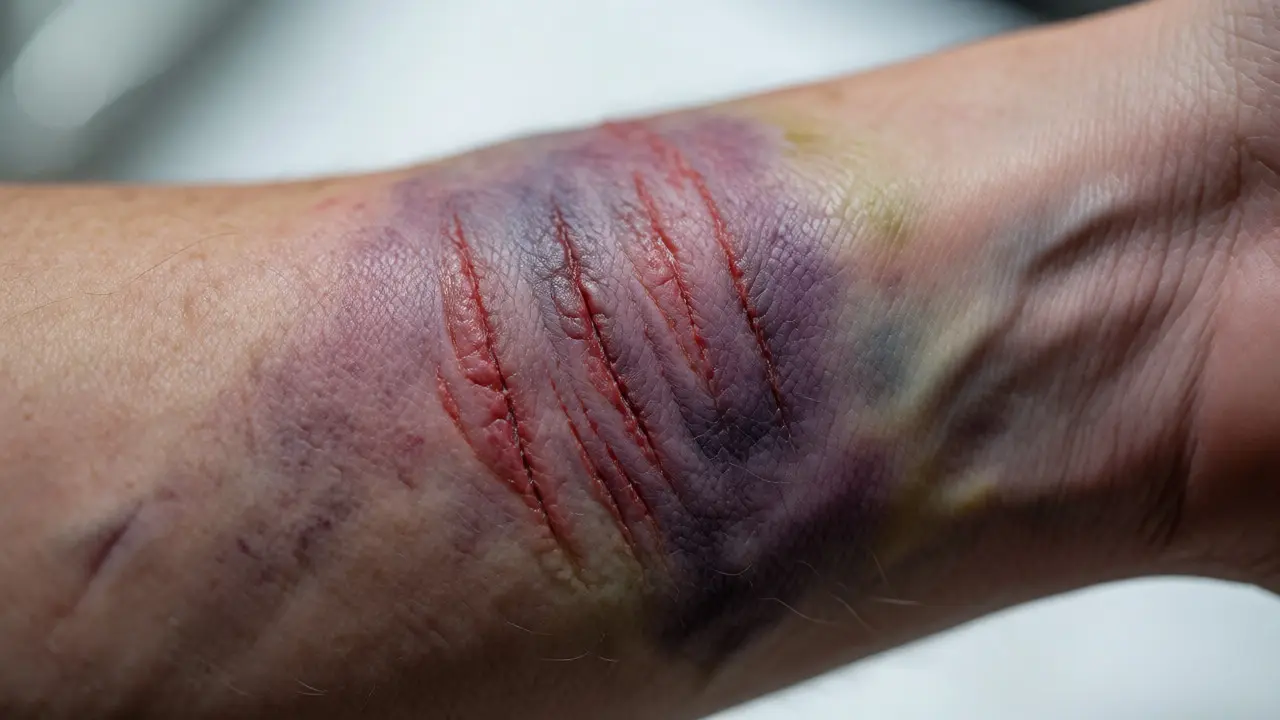
You walk into a room. Someone falls. Or maybe they don't fall-maybe someone hits them with a baseball bat, or a car bumper clips their leg at thirty miles per hour. The aftermath looks chaotic to the untrained eye: purple skin, broken bones, swelling. But to a forensic pathologist, that chaos is a language. Blunt force trauma is mechanical injury caused by non-penetrating impact from an object or surface, leaving behind specific signatures on bone and tissue. Understanding these signatures isn't just about naming the injury; it's about reconstructing the event. Who held the weapon? What angle did it hit? Was the person standing or lying down when the blow landed?
The Physics of Impact: Why Bones Break the Way They Do
Bones aren't uniform sticks. They are composite materials, strong in compression but weaker in tension and shear. When Blunt Force Trauma hits the body, the outcome depends entirely on three variables: the mass of the object, the velocity of the impact, and the area over which that force is distributed. A hammer delivers concentrated energy; a steering wheel spreads it out. This distinction dictates whether you see a clean crack or a shattered mess.
Think about a green branch versus a dry one. Young people have more elastic bone matrix, meaning their bones bend before they break. Elderly individuals often suffer comminuted fractures (shattered into multiple pieces) because osteoporosis has stripped away that elasticity. In forensic analysis, we look for the fracture line's direction. If a long bone like the femur snaps in a spiral pattern, it usually suggests torsion-a twisting force, perhaps from a fall where the foot caught on pavement while the body kept moving. A transverse fracture (straight across) typically indicates a direct, perpendicular blow.
We also categorize fractures based on how the bone failed under stress:
- Linear Fractures: Simple cracks without displacement. Common in skull injuries from moderate impacts.
- Comminuted Fractures: The bone breaks into three or more fragments. This requires high-energy transfer, like a high-speed vehicle collision.
- Depressed Fractures: Specifically seen in the skull when a heavy object strikes with enough force to push bone inward toward the brain.
- Greenstick Fractures: One side of the bone bends and cracks, while the other remains intact. Almost exclusively found in children due to softer bone density.
Bruising Mechanics: Reading the Color Code of Time
If fractures tell us about structural failure, bruises (contusions) tell us about vascular damage. A bruise forms when capillaries rupture under blunt impact, leaking blood into surrounding soft tissue. The tricky part? The color changes over time as hemoglobin-the oxygen-carrying protein in red blood cells-breaks down. Forensic experts use this degradation timeline to estimate how long ago the injury occurred, though it’s far from perfect science.
Here’s the typical progression of a superficial bruise in fair-skinned individuals:
- 0-12 Hours: Red or purple. Fresh oxygenated blood leaks out.
- 12-48 Hours: Blue or dark purple. Oxygen is consumed; deoxyhemoglobin takes over.
- 5-7 Days: Green. Hemoglobin breaks down into biliverdin.
- 7-10 Days: Yellow/Brown. Biliverdin converts to bilirubin and then hemosiderin.
- 2+ Weeks: Fades to light brown or disappears entirely.
But here’s the catch: depth matters. Deep muscle bruises might not show up on the skin surface for days, appearing suddenly as if from nowhere. Conversely, facial bruises can spread rapidly due to gravity and loose tissue planes, making the initial site of impact hard to pinpoint. If you see bruising around the eyes (raccoon eyes) after a head injury, it often signals a basilar skull fracture, where blood tracks along the anterior cranial fossa. It’s not necessarily a punch to the face; it’s internal leakage finding its way to the surface.
Patterned Injuries: The Object Leaves Its Mark
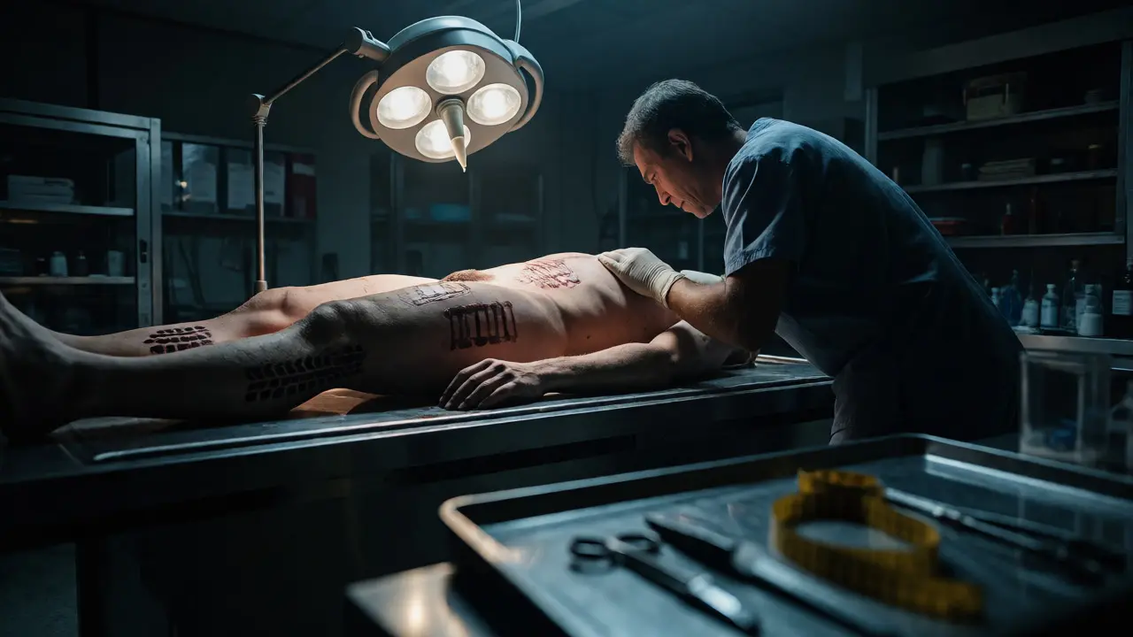
Not all blunt force trauma results in generic swelling. Sometimes, the object striking the body leaves a distinct imprint, known as a patterned injury. This is gold for investigators trying to match a suspect’s weapon to the victim’s wounds.
Consider a belt buckle. If struck against the back, it leaves a rectangular abrasion with a central depression matching the buckle’s shape. A tire tread mark on a pedestrian’s leg mirrors the rubber pattern exactly. Even clothing can act as a stencil; a zipper pressed against skin during a struggle will leave a linear abrasion that mimics the teeth of the fastener.
We classify these marks into two main types:
| Feature | Abrasion (Scrape) | Contusion (Bruise) |
|---|---|---|
| Mechanism | Tangential friction strips epidermis. | Perpendicular crush ruptures vessels. |
| Appearance | Reddened, raw, sometimes scabbed. | Purple/blue/yellow discoloration beneath skin. |
| Pattern Retention | High fidelity; retains object texture well. | Lower fidelity; diffuse edges blur details. |
| Healing Time | Days to weeks depending on depth. | Weeks to months for full resolution. |
One common misconception is that "slap marks" always look like hands. In reality, hand bruises are rarely clear imprints of fingers. The palm spreads out upon impact, creating a broad, oval-shaped bruise. Finger pads might leave small circular subcutaneous hemorrhages, but unless the skin is very thin (like on the eyelids), you won’t see a neat fingerprint. Always look for defensive wounds on the forearms-these are often mixed abrasions and fractures from blocking blows.
Skull Fractures: Distinguishing Antemortem from Postmortem
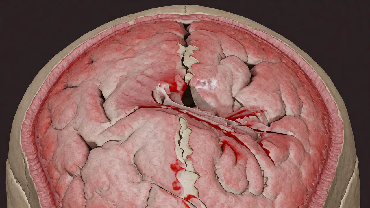
The skull is the most scrutinized bone in forensic cases because it protects the brain, and damage here is frequently fatal. However, distinguishing between a fracture sustained while alive (antemortem) and one caused after death (postmortem) is critical. After death, tissues lose turgor pressure. The scalp sags, and the bone becomes brittle. A postmortem drop onto a hard floor can shatter the skull easily, mimicking a severe beating.
How do we tell the difference? We look for vital reactions. Antemortem fractures show bleeding within the bone layers (diploë) and swelling of the dura mater. There may be clotting in the epidural space. Postmortem fractures lack these biological responses; the edges are dry, and there is no inflammation. Additionally, antemortem fractures often radiate outward from the point of impact, following lines of weakness in the cranial sutures. Postmortem fractures tend to be irregular and localized to the contact point.
Another key indicator is the presence of bridging veins. Subdural hematomas occur when these veins tear, often due to acceleration-deceleration forces (like shaking a baby or a whiplash-style car crash). Unlike epidural hematomas, which are usually arterial and form lens-shaped clots, subdural bleeds crescent-shaped and spread widely. Identifying the type of hematoma helps determine if the cause was a single heavy blow or repeated minor impacts.
Internal Organs: The Hidden Damage
Skin tells half the story. The other half lies inside. Blunt force trauma doesn’t need to break the skin to kill. Liver and spleen lacerations are among the most common causes of death from abdominal blunt trauma. These organs are solid, highly vascular, and relatively fragile. A seatbelt injury during a car accident can compress the abdomen against the spine, shearing the liver.
Pathologists examine organ capsules for tears. A healthy capsule is smooth and shiny. A ruptured one reveals jagged edges and active bleeding. Rib fractures often accompany these injuries, especially if the ribs were fractured inward, piercing the lung or heart. However, elderly patients may have rib fractures without significant internal damage due to calcified cartilage absorbing some shock, whereas younger patients might sustain severe organ damage with fewer visible external signs.
Always check the kidneys. They are retroperitoneal (located behind the abdominal cavity), so they’re somewhat protected, but a direct kick to the flank can cause hematuria (blood in urine) and renal contusion. In cases of suspected abuse, bilateral kidney injury is a red flag, suggesting systematic rather than accidental harm.
Contextualizing the Evidence: Accident, Suicide, or Homicide?
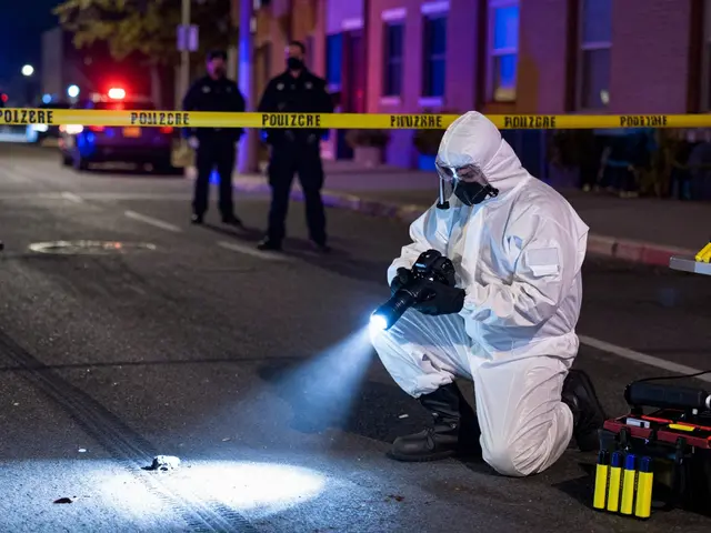
Finally, we must interpret the findings within the scene context. A single depressed skull fracture on the top of the head could be a fall from height, a self-inflicted blow with a hammer, or an attack from above. How do we differentiate?
We look at distribution. Homicidal beatings often involve multiple weapons and varied angles. You might find defensive fractures on the ulna (forearm bone) alongside blunt trauma to the parietal bone. Suicidal blunt force trauma is rare but possible; it usually involves a single, massive impact to the forehead or temple, often with hesitation marks (minor prior attempts) nearby. Accidental trauma tends to be isolated to areas exposed during a fall, such as knees, elbows, and the occipital region (back of the head).
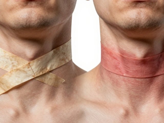
Remember, no single piece of evidence is conclusive in isolation. A bruise on the neck could mean strangulation, or it could mean a tight collar. A fractured hyoid bone suggests manual compression, but it can also happen in hanging deaths. The job of the forensic analyst is to weave the physical evidence-fractures, bruises, internal bleeding-into a coherent narrative that aligns with the physics of the scene and the biology of the victim.
Can bruising appear hours after an injury?
Yes. Deep tissue bruises often take 12 to 24 hours to become visible on the skin surface. Blood from deeper vessels migrates slowly through tissue planes before reaching the dermis. This delay can make initial examinations misleading if the patient is assessed immediately after the incident.
Do all fractures indicate blunt force trauma?
No. Pathological fractures can occur in weakened bones (due to cancer, osteoporosis, or infection) with minimal or no trauma. Additionally, spontaneous fractures can happen during normal movement in severely compromised bone structures. Forensic analysis checks for underlying disease markers in the bone sample.
How accurate is dating a bruise by color?
It is an estimation, not a precise clock. Factors like age, medication (blood thinners accelerate bruising), lighting conditions, and skin tone significantly affect visibility. Forensic pathologists use color as a rough guide combined with other histological findings, never relying on it alone for exact timing.
What is a "Tardieu spot"?
A Tardieu spot is a small, pinpoint hemorrhage found in the lungs, heart, or conjunctivae (eyes) resulting from intense thoracic compression. While historically associated with suffocation or strangulation, they can also appear in sudden cardiac arrest or severe chest trauma, so they are supportive but not diagnostic on their own.
Can a fall from standing height cause a skull fracture?
Yes, particularly in the elderly or those with anticoagulant therapy. While less common in young adults, a direct impact on a hard surface like concrete can generate sufficient force to fracture the temporal or parietal bones, especially if the head strikes a corner or edge.