You walk into a room and see a dark patch on the floor. Is it a spill? A stain? Or evidence of something more serious? In the world of forensic science, specifically within the study of physical evidence at crime scenes, that question isn't just about color. It's about volume. When we talk about "saturation" in this context, we aren't discussing oxygen levels or iron content in the bloodstream. We are asking a very specific, practical question: how much blood has actually soaked into a material, and what does that tell us about the event?
This is where Bloodstain Pattern Analysis (BPA) comes in. BPA is the scientific discipline used to interpret the shape, size, and distribution of blood stains. One of the critical components of this analysis is understanding saturation-the point at which a surface can hold no more liquid. If you pour a cup of water onto a dry sponge, it absorbs quickly. Keep pouring, and eventually, the sponge becomes saturated, and the water starts pooling on top. Blood behaves similarly, but with far more complex variables like viscosity, surface tension, and the porous nature of the substrate.
The Science of Absorption and Saturation
To understand how much blood remains on a surface, you first need to understand the difference between porous and non-porous materials. This distinction dictates everything from how the blood looks to how much of it stays put versus how much spreads out.
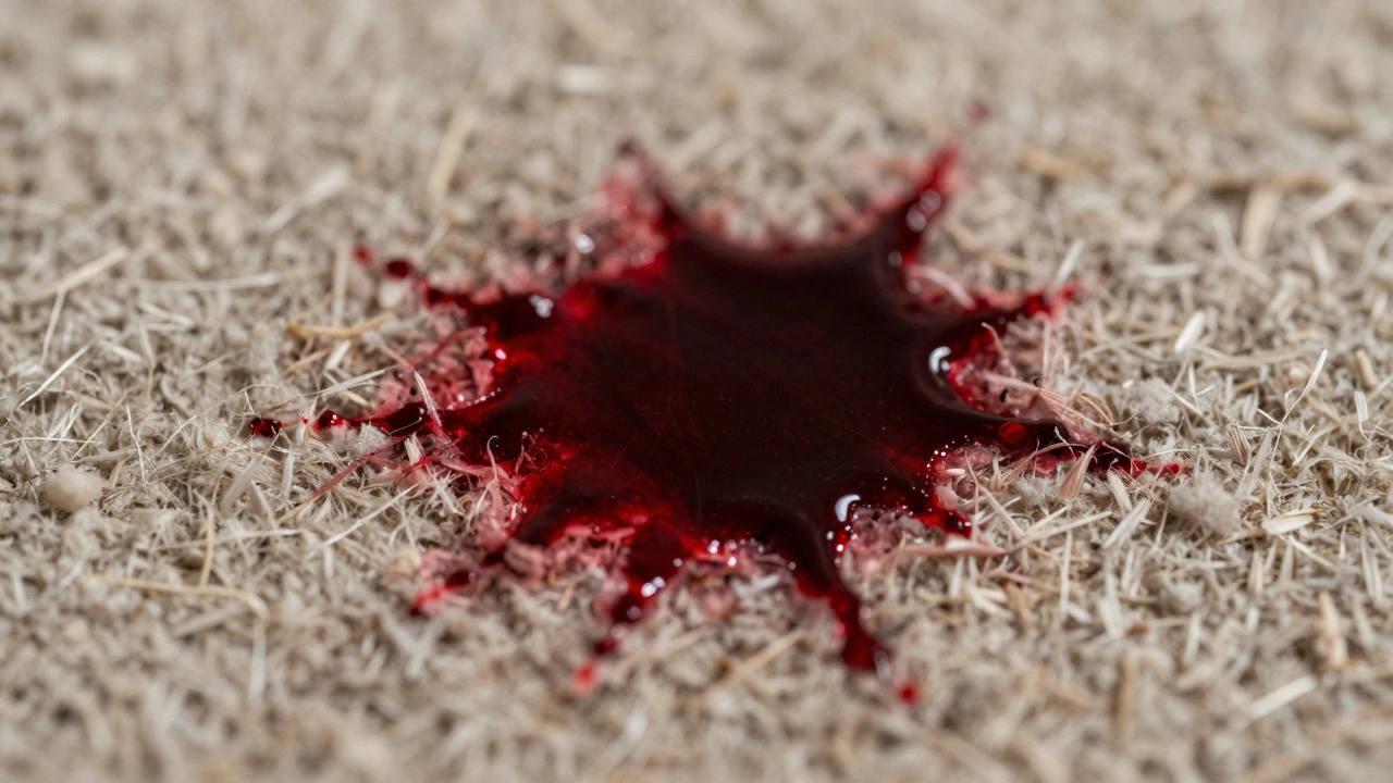
Porous surfaces, such as carpet, wood, fabric, and paper, absorb blood through capillary action. The liquid is drawn into the microscopic gaps between fibers or pores. On these surfaces, saturation happens relatively quickly depending on the thickness of the material. Once the internal structure is full, the blood may begin to wick outward, creating larger, diffuse stains rather than distinct drops. The amount of blood "on" the surface is often less visible than the amount "in" the surface. This makes quantifying the original volume difficult because the blood disappears into the matrix of the material.
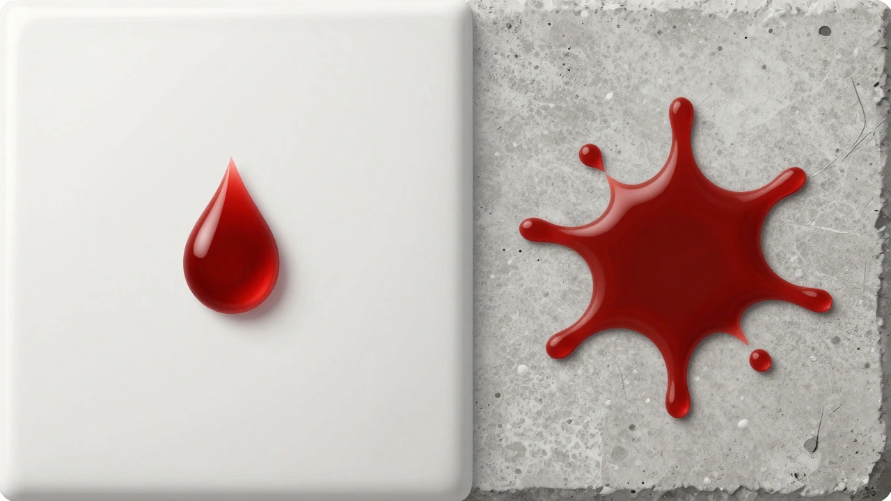
In contrast, non-porous surfaces like glass, tile, metal, or plastic do not absorb blood. The blood sits on top. Here, saturation refers to the volume required to cover a specific area completely. Because there is no absorption, the blood maintains its three-dimensional shape until it dries or is disturbed. This allows investigators to measure height, width, and volume with much greater precision. A single drop on a tile floor tells a different story than a single drop on a wool rug, even if the initial volume was identical.
Factors Influencing Blood Volume on Surfaces
It isn't just about the floor type. Several dynamic factors influence how much blood ends up on a surface and how it presents itself.
- Viscosity: Fresh blood is thicker than diluted blood. If blood mixes with water or another fluid, its viscosity decreases, causing it to spread further and saturate a larger area before drying. High-viscosity blood tends to stay more localized.
- Surface Texture: Even on non-porous surfaces, texture matters. A rough concrete floor will hold more blood in its crevices than a smooth linoleum sheet. The "contact angle"-how sharply the blood bead sits against the surface-changes based on cleanliness and texture.
- Gravity and Angle: Blood on a vertical wall behaves differently than on a horizontal floor. Gravity pulls the blood downward, causing it to run or drip. This creates "flow patterns" that can indicate the orientation of the object when the bleeding occurred.
- Drying Time: As blood dries, it loses moisture. The remaining hemoglobin and cellular material shrink and darken. A wet stain covers more area than a dried one. Understanding the stage of drying helps estimate the time since deposition.
Quantifying the Stain: From Visuals to Volume
How do investigators actually determine "how much" blood is there? They don't usually weigh the stain directly at the scene. Instead, they use geometric calculations and comparative analysis.
For individual droplets on non-porous surfaces, analysts can estimate the volume based on the diameter of the stain. A standard human blood drop is roughly spherical when falling. When it hits a flat, non-absorbent surface, it spreads out. By measuring the diameter of the resulting circular stain, experts can reverse-calculate the approximate volume of the drop. Small drops might be 0.05 milliliters, while larger, forceful impacts can produce drops exceeding 0.1 milliliters. Multiply that by the number of drops, and you get a total volume estimate.
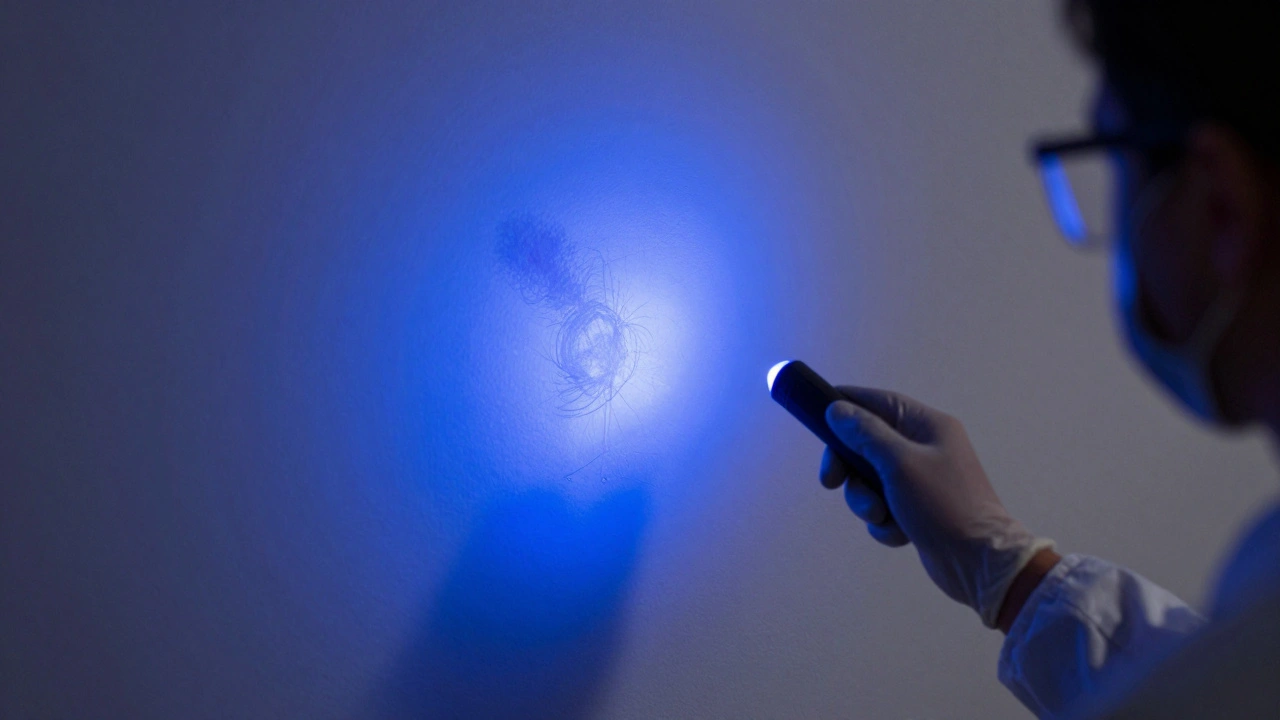
On porous surfaces, this calculation is trickier. Investigators look for "saturation zones." If a stain appears uniformly dark and wet-looking in the center, that area is likely saturated. The edges, where the color fades, indicate where the capillary action stopped. Chemical tests, such as luminol or Bluestar, can reveal the true extent of the stain, especially if the blood has been cleaned. These chemicals react with the iron in hemoglobin, glowing under specific light conditions, revealing the total area affected regardless of visibility.
| Surface Type | Absorption Rate | Saturation Indicator | Volume Estimation Difficulty |
|---|---|---|---|
| Ceramic Tile | None | Pooling or beading | Low (High Precision) |
| Carpet | High | Wicking and darkening | High (Low Precision) |
| Wood | Moderate | Penetration depth | Moderate |
| Glass | None | Surface tension beads | Low (High Precision) |
The Role of Hematocrit in Saturation
Here is a detail that often surprises people: not all blood is the same. The hematocrit level-the percentage of red blood cells in the blood-affects how blood interacts with surfaces. Individuals with higher hematocrit levels have thicker, more viscous blood. This blood doesn't spread as easily as blood from someone with lower hematocrit. It tends to form taller, narrower spatter patterns and saturates surfaces more slowly. Conversely, diluted blood (perhaps mixed with sweat or rainwater) spreads faster and covers a wider area with less volume. Analysts must account for these biological variations when interpreting saturation patterns.
Why Saturation Matters in Investigations
Understanding saturation isn't just an academic exercise; it changes the narrative of a crime scene. Consider a scenario where a witness claims a victim was standing still while bleeding. If the blood on the floor shows signs of high saturation and pooling, it suggests the person remained in one place for a significant time, allowing gravity and absorption to take effect. If the blood is scattered in small, unsaturated droplets, it indicates movement, struggle, or rapid evacuation.
Saturation also helps distinguish between primary and secondary transfer. Primary transfer occurs when blood moves directly from a source to a surface. Secondary transfer happens when a bloody object touches another surface. Secondary stains are often lighter, less saturated, and irregular because only a fraction of the original blood is transferred. Recognizing this difference prevents investigators from misinterpreting the severity or location of the injury.
Challenges in Modern Crime Scene Analysis
Modern cleaning products pose a unique challenge to saturation analysis. Many households use enzymatic cleaners designed to break down blood proteins. These cleaners can remove the visible stain without removing the chemical signature detectable by luminol. However, they can alter the surface's porosity. A treated carpet might absorb less blood in subsequent incidents, changing the saturation dynamics. Investigators must document the condition of the scene meticulously, noting any signs of cleaning attempts, as this affects the reliability of volume estimates.
Additionally, environmental factors like temperature and humidity play a role. In hot, dry environments, blood evaporates faster, reducing the apparent volume and altering the saturation pattern before it can be fully documented. In humid conditions, blood takes longer to dry, increasing the risk of accidental smearing or spreading due to gravity, which can distort the original saturation footprint.
Practical Steps for Documenting Saturation
If you are involved in documenting a scene, whether as a professional or for educational purposes, follow these steps to capture saturation data accurately:
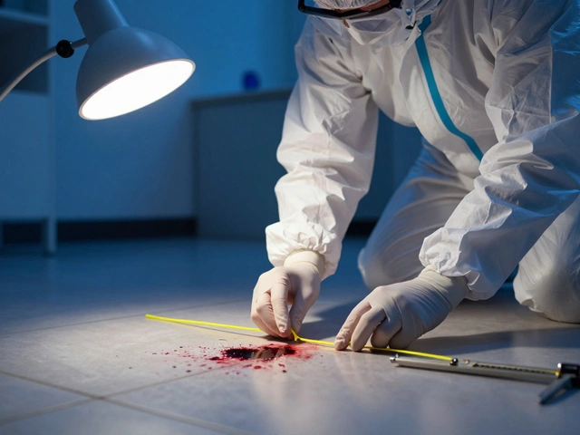
- Photograph Immediately: Take close-up photos of the stain with a scale ruler. Capture both the overall area and detailed textures. Use cross-polarized lighting to reduce glare on non-porous surfaces.
- Note Surface Conditions: Record whether the surface is wet, dry, clean, or dirty. Note the material type explicitly.
- Assess Color Variation: Look for darker centers (saturation zones) versus lighter edges (wicking zones). This gradient provides clues about the volume and direction of flow.
- Test for Latent Blood: Use presumptive tests like Kastle-Meyer or luminol to reveal hidden areas. Remember, these tests show presence, not necessarily volume, but they define the total boundary of the saturation event.
- Preserve Samples: Collect samples from both saturated and unsaturated areas for laboratory comparison. This helps establish baseline absorption rates for that specific material.
By focusing on the physical interaction between blood and surfaces, we move beyond guesswork. We build a factual record of what happened, grounded in the physics of fluids and the chemistry of biology. Saturation is the silent witness, telling us not just where the blood went, but how much was there to begin with.
What is the difference between blood absorption and saturation?
Absorption is the process by which a porous material takes in blood. Saturation is the point at which the material can no longer absorb any more liquid. Once saturated, additional blood will pool on the surface or spread laterally rather than penetrating deeper into the material.
How does hematocrit affect bloodstain patterns?
Hematocrit is the ratio of red blood cells to plasma. Higher hematocrit means thicker, more viscous blood. This results in smaller, rounder stains that do not spread as far. Lower hematocrit leads to thinner blood that spreads more easily, creating larger, flatter stains with the same volume.
Can you estimate the volume of blood from a stain on carpet?
Estimating exact volume on carpet is difficult due to absorption and wicking. However, analysts can estimate relative volume by comparing the size and darkness of the stain to known standards. Larger, darker stains generally indicate higher volumes, but precise measurement requires controlled experiments with similar carpet types.
Why do blood stains change color over time?
Fresh blood is bright red due to oxygenated hemoglobin. As it dries and oxidizes, it turns brown, then black. The concentration of hemoglobin increases as water evaporates, making the stain appear darker and more concentrated in the center, which helps identify saturation zones.
Does cleaning a surface remove all evidence of blood saturation?
Cleaning removes visible blood but rarely eliminates all traces. Enzymatic cleaners break down proteins, but chemical residues remain. Tests like luminol can detect blood even after thorough cleaning. However, cleaning alters the surface's porosity, which can complicate future saturation analysis.