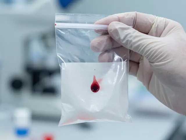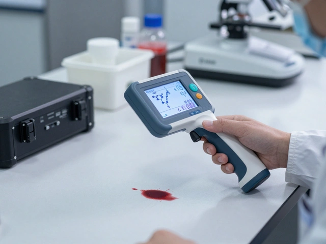
Imagine walking into a room where the walls are streaked with red. Most people see chaos. A forensic expert sees physics. Specifically, they see the pressure behind the blood. When an artery is cut, it doesn't just drip; it pulses. This creates a specific type of arterial spray that tells investigators exactly how the injury happened and when. Unlike passive drips or gravity-driven flows, this pattern is dynamic, directional, and often dramatic. Understanding it is the difference between guessing a timeline and proving it.
What Defines Arterial Spray?
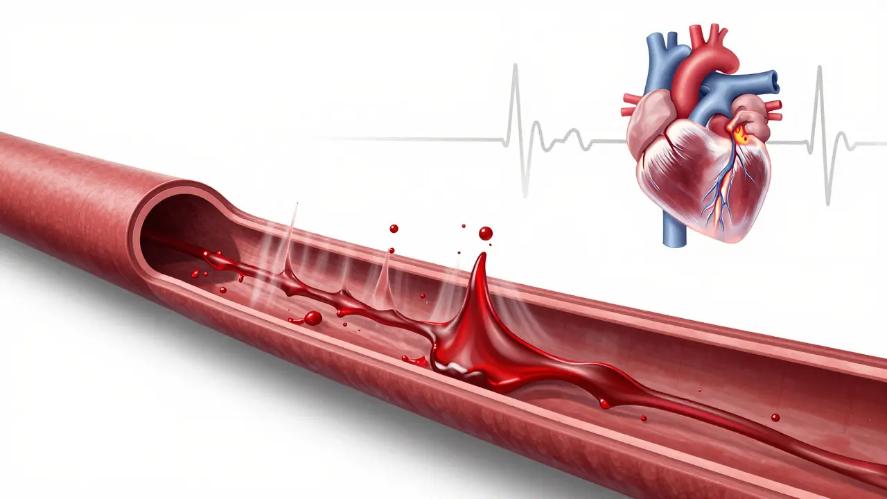
Arterial spray is a bloodstain pattern produced by blood being expelled from a severed or damaged artery under pulsatile pressure. To understand why it looks the way it does, you have to look at human physiology. Your heart pumps blood through arteries in rhythmic contractions known as systole (contraction) and diastole (relaxation). When an artery like the femoral or carotid is breached, the blood shoots out during each beat of the heart. It doesn't flow steadily; it spurts.
This mechanism creates a distinct visual signature. You will typically see a fine mist of small droplets mixed with larger drops. The pattern often radiates outward from the point of impact. If the victim was moving, the spray follows their path. If they were still, it projects forward based on the angle of the wound. The key attribute here is pulsatility. In a live person, the spray comes in bursts corresponding to the heartbeat. Once death occurs, the heart stops, and the spray ceases immediately, leaving behind a static record of the final moments.
The Physics of Blood Ejection
Blood is not just liquid; it has viscosity and momentum. When an artery ruptures, the pressure can range from 120 mmHg (systolic) down to 80 mmHg (diastolic) in a healthy adult. However, in trauma cases, shock and blood loss lower this pressure over time. Forensic scientists use the size and distribution of the droplets to estimate the energy involved.
- Fine Mist: Indicates high-energy ejection, often seen when the artery is fully severed and unobstructed.
- Larger Droplets: May indicate partial obstruction or lower pressure as circulation fails.
- Directionality: The tail of the elongated stains points in the direction of travel. This helps reconstruct the movement of the victim or the weapon.
This physical behavior distinguishes arterial spray from other patterns. For instance, a venous bleed produces a steady flow or drip because veins operate under much lower pressure (typically less than 15 mmHg). An arterial bleed, by contrast, is explosive. The distinction is critical for determining whether a wound was inflicted while the victim was alive and conscious.
Distinguishing Arterial Spray from Other Patterns
Mistaking one bloodstain pattern for another can derail an investigation. Let's compare the three primary types of active bleeding patterns: passive, arterial, and impact.
| Pattern Type | Source Pressure | Visual Characteristics | Typical Wound Source |
|---|---|---|---|
| Passive Flow | Gravity / Low Venous | Straight lines, pooling, dripping | Cut vein, capillary bed |
| Arterial Spray | Pulsatile Arterial (High) | Fine mist, directional streaks, spatter clusters | Severed artery (e.g., femoral, carotid) |
| Impact Spatter | External Force | Radiating from impact point, varied sizes | Blunt force trauma, gunshot |
Notice the difference in the "Visual Characteristics" column. Passive flow is predictable and vertical. Impact spatter radiates from a single point of contact. Arterial spray, however, is unique because it combines fine mist with directional projection. It often looks like a fan or a cone extending from the wound site. If you see a wall covered in tiny dots that get larger as they move away from a central source, you are likely looking at arterial spray.
Reconstructing the Crime Scene
Why do investigators care about this? Because arterial spray is a clock. Since the pattern depends on a beating heart, its presence confirms the victim was alive at the moment of injury. More importantly, the length and intensity of the spray tell you how long the bleeding lasted before the heart stopped.
In a case study from Portland, Oregon, investigators analyzed a scene where a victim was found with a deep laceration to the neck. The arterial spray extended six feet up the opposite wall. By measuring the height and density of the droplets, experts determined the victim had been standing upright for approximately 45 seconds after the initial strike. This detail helped rule out a staged scene where the body might have been moved after death. If the body had been moved post-mortem, the spray would have stopped abruptly without the characteristic tapering seen in natural cessation.
Investigators also look for "satellite spatter." These are secondary droplets that bounce off the primary spray and land further away. Their presence indicates a high-velocity event, reinforcing the theory of a severe arterial breach rather than a minor cut.
Factors That Alter the Pattern
Real-world scenes are messy. Several variables can distort the classic arterial spray pattern, making interpretation tricky. Understanding these factors prevents false conclusions.
- Body Position: If the victim falls, the spray changes direction. A horizontal body produces a different trajectory than an upright one. Investigators must map the entire floor and walls to track these shifts.
- Obstruction: Clothing, bandages, or even the victim's own hand pressing against the wound can partially block the spray. This results in a narrower, more concentrated stream rather than a wide fan.
- Environmental Conditions: Wind or air conditioning can blow fine mist droplets sideways. In outdoor scenes, rain can wash away smaller droplets, leaving only the larger, heavier drops. This can make the pattern look less intense than it actually was.
- Time of Death: As blood pressure drops due to hypovolemic shock, the spray becomes weaker. The final inches of the pattern may show fewer droplets and less reach. This "fading" effect is crucial for estimating the time of death relative to the injury.
For example, if you find a dense cluster of spray near the body but a sparse trail leading to the door, it suggests the victim walked toward the exit while losing strength. The transition from dense to sparse is a direct reflection of physiological decline.

Common Misinterpretations
Even experienced examiners can make mistakes. One common error is confusing arterial spray with shotgun spatter. Both produce fine mist and directional patterns. However, shotgun spatter originates from a projectile, so it radiates from the entry wound in a star-like pattern. Arterial spray originates from the vessel itself, so it projects away from the wound in a stream or fan. Looking at the wound location relative to the stain origin solves this puzzle.
Another mistake is assuming all fine mist is arterial. High-velocity impact from a hammer or axe can create similar fine droplets. The key differentiator is the consistency. Arterial spray maintains a rhythmic quality in its distribution (though hard to see statically), while impact spatter is random and chaotic. Additionally, arterial spray often shows a clear relationship to the anatomical location of major arteries (neck, groin, armpit), whereas impact spatter can occur anywhere on the body.
Practical Tips for Documentation
If you are documenting a scene with potential arterial spray, precision is everything. Here is how professionals handle it:
- Photograph Before Touching: Use a scale ruler in every photo. Capture wide shots for context and close-ups for droplet size.
- Map the Trajectory: Draw arrows indicating the direction of the tails. This shows the vector of the blood ejection.
- Note Surface Texture: Smooth walls allow blood to spread; rough surfaces cause splashing. Note the material of every surface involved.
- Collect Samples: Take samples from the center of the spray (highest concentration) and the edges (lowest concentration). This helps lab technicians analyze the degradation of the blood cells over distance.
These steps ensure that the data collected in the field supports the laboratory analysis later. Without proper documentation, the story the blood tells gets lost in translation.
Frequently Asked Questions
How far can arterial spray travel?
In rare cases with full arterial severance and high blood pressure, fine mist can travel over 20 feet. However, most significant staining occurs within 5 to 10 feet of the source. The distance depends heavily on the angle of the wound and the victim's position.
Does arterial spray stop immediately upon death?
Yes. Arterial spray is driven by cardiac contraction. Once the heart stops, the pulsatile ejection ceases instantly. Any remaining blood will then flow passively due to gravity, creating a different, non-pulsatile pattern.
Can you tell which artery was cut from the pattern alone?
Not definitively from the pattern alone, but the location of the spray relative to the body provides strong clues. Spray from the neck projects upward and backward; spray from the femoral artery projects downward and forward. Combining this with autopsy findings confirms the specific vessel.
How does temperature affect arterial spray patterns?
Temperature affects blood viscosity slightly, but its bigger impact is on preservation. Cold environments preserve fine mist droplets better, allowing for more detailed analysis. Hot environments can cause rapid drying, which alters the shape of the stains and makes measurement difficult.
Is arterial spray always visible to the naked eye?
The larger droplets are usually visible. However, the finest mist particles may be too small to see clearly without magnification or alternate light sources. UV light can help reveal dried micro-droplets that are invisible under standard lighting.