You’ve seen the movies. The body is on the table, the lights are bright, and a doctor in scrubs makes a quick cut to find the bullet or the poison. But real forensic pathology isn’t about rushing to the internal organs. It starts long before the scalpel touches skin. It starts with the external examination, a meticulous, methodical review of the body’s surface that often tells the whole story.
If you’re curious about how investigators determine what happened to a person after they died, understanding this first step is crucial. It’s not just about looking for wounds; it’s about reading the body’s final moments like a map. This process answers critical questions: Was the body moved? Did the person struggle? When did they die? Let’s break down exactly what pathologists look for and why every inch of skin matters.
The Big Picture: Why Start Outside?
Think of the external exam as gathering the clues before opening the box. Once you start cutting, you change the body’s state. You drain fluids, remove organs, and alter tissues. If you miss something on the outside, you might lose it forever. That’s why the National Association of Medical Examiners (NAME) guidelines stress thoroughness here. A missed ligature mark on the neck can turn a homicide into an accident. A faint bruise on the back can reveal if someone was struck while lying down.
This phase is also where context meets biology. The pathologist doesn’t work in a vacuum. They have police reports, scene photos, and witness statements. The external exam validates or contradicts these narratives. If a suspect claims the victim fell forward, but there are defensive wounds on the hands and bruising on the shins, the story changes fast.
Reading the Skin: Lividity and Rigor
Two of the earliest indicators of time since death are livor mortis (lividity) and rigor mortis (rigor). These aren’t perfect clocks, but they give a rough timeline when combined with other data.
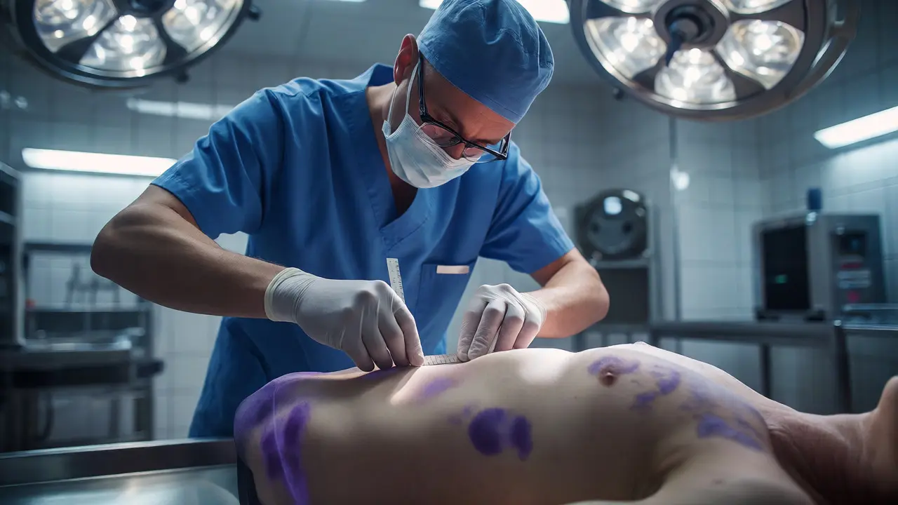
Lividity happens because blood stops circulating and settles due to gravity. It appears as purple-red discoloration in the lowest parts of the body. Here’s the catch: lividity becomes "fixed" after about 8-12 hours. Before that, if you press on the skin, the color blanches (turns white). After it fixes, it stays put. This helps answer a huge question: Was the body moved? If you find lividity on the back of a body found face-down, the person died in one position and was flipped later. That’s a major red flag for foul play or staging.
Rigor mortis is the stiffening of muscles due to chemical changes in muscle fibers. It typically begins in small muscles (like the jaw) within 2-4 hours, spreads to the rest of the body over 6-12 hours, and then fades over the next 24-36 hours. Pathologists check the jaw, arms, and legs to gauge where the body is in this cycle. It’s tricky because temperature affects it-heat speeds it up, cold slows it down-but it’s a vital piece of the puzzle.
Identifying Trauma: Wounds vs. Artifacts
Not all marks on a body are injuries. Some happen during transport, some from medical attempts to save life, and some from post-mortem handling. Distinguishing antemortem injuries (those sustained while alive) from postmortem artifacts is a core skill.
Antemortem wounds bleed. Even minor cuts will show clotting or inflammation if the heart was still pumping. Postmortem cuts don’t bleed significantly because circulation has stopped. Pathologists look for "vital reaction"-swelling, redness, or cellular changes around a wound. If a bruise has no swelling, it likely happened after death.
Common trauma types include:
- Abrasions: Scrapes where the top layer of skin is removed. Directionality can show which way the force came from.
- Contusions: Bruises caused by broken blood vessels under the skin. Their color changes over time, helping estimate age.
- Lacerations: Tearing of tissue, often from blunt force. Unlike sharp-force cuts, lacerations have "bridging strands" of tissue across the wound.
- Incised wounds: Clean cuts from sharp objects like knives or glass.
Specific Clues: Hands, Neck, and Genitals
Certain areas get extra attention because they tell specific stories.
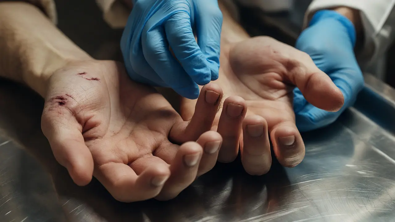
The Hands: Check the palms and backs of hands. Defensive wounds-cuts on the palm from grabbing a knife-are classic signs of struggle. Also, look under the fingernails. Scraps of skin or fabric trapped there can identify an attacker through DNA analysis. This is often the single most valuable piece of physical evidence collected during an autopsy.
The Neck: In cases of hanging or strangulation, the neck holds the key. Ligature marks (from ropes or cords) have distinct patterns. Hanging usually leaves a V-shaped mark that goes up toward the ears. Strangulation often leaves a horizontal band. Petechial hemorrhages-tiny pinpoint bleeds in the eyes, eyelids, or face-suggest pressure was applied to the neck, blocking blood flow out of the head.
Genitals: In sexual assault cases, swabs are taken here before any washing occurs. Presence of semen, hair, or foreign materials is documented. Even in non-sexual deaths, genital inspection can reveal injuries consistent with abuse or accidents.
Documentation: Photos, X-Rays, and Swabs
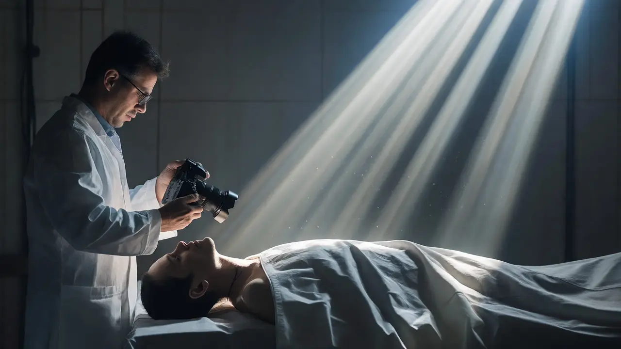
Memory is fallible. Photos are permanent. Every significant finding gets photographed with a scale ruler for size reference. Close-ups show texture; wide shots show location relative to anatomical landmarks (like the nipple line or navel).
X-rays are standard practice now. They reveal hidden fractures, bullets, or shrapnel without cutting into the body. A bullet fragment lodged deep in muscle might be missed visually but shows up clearly on an X-ray. This guides the pathologist on where to dissect internally.
Swabbing is another critical step. Before cleaning the body, pathologists swab potential trace evidence: dirt under nails, fibers on clothing, saliva on bite marks. These samples go straight to the lab for toxicology or DNA testing. Once the body is washed, those clues are gone.
| Indicator | What It Is | Why It Matters |
|---|---|---|
| Livor Mortis | Blood pooling due to gravity | Determines if body was moved; estimates time since death |
| Rigor Mortis | Muscle stiffening | Helps narrow down time of death window |
| Petechiae | Tiny eye/skin bleeds | Suggests suffocation or high-pressure trauma |
| Defensive Wounds | Injuries on hands/arms | Indicates victim fought back |
| Tattoo Scarring | Healed ink deposits | Confirms identity when faces are damaged |
When the External Exam Solves the Case
Sometimes, the internal exam confirms what the external already proved. Other times, the external findings are enough to close the case. If a body shows clear signs of advanced decomposition, massive trauma incompatible with survival, or obvious disease markers (like severe jaundice), the pathologist might decide an full internal dissection isn’t necessary. This saves resources and respects the deceased.
But even if the cause of death seems obvious, the external exam remains mandatory. It provides the baseline for legal testimony. Defense attorneys will scrutinize whether every detail was recorded. Missing photos or skipped swabs can create reasonable doubt. Precision here protects the integrity of the entire investigation.
For anyone interested in criminal justice or medicine, remember this: the dead do speak, but only if we listen carefully to their skin. The external examination is that act of listening. It turns silent bodies into witnesses.
How long does an external examination take?
It varies widely. A straightforward case might take 30-60 minutes. Complex cases with multiple injuries, extensive documentation needs, or unknown identities can take several hours. There is no rush; accuracy trumps speed.
Can livor mortis be mistaken for bruising?
Yes, especially in darker-skinned individuals. Pathologists differentiate them by pressing the area. Lividity blanches (whitens) under pressure if not fixed; bruises generally do not. Location also helps-lividity follows gravity, bruises follow impact sites.
Do forensic pathologists wear gloves during the external exam?
Always. Gloves protect the examiner from pathogens and prevent contamination of the evidence. Changing gloves between different body regions or tasks is standard protocol to avoid cross-contamination of DNA or trace evidence.
What happens if the body is too decomposed for an external exam?
Pathologists still perform it. They may clean the body gently to expose features. Decomposition masks some details but reveals others, like insect activity which helps estimate post-mortem interval. Bones and teeth often remain identifiable even when soft tissue is gone.
Is an external exam the same as a visual inspection at the crime scene?
No. Crime scene inspections are preliminary and done by detectives or medical examiners on-site. The formal external examination happens in the morgue under controlled lighting and conditions, allowing for detailed measurements, photography, and sample collection that aren’t possible outdoors.