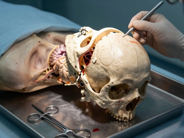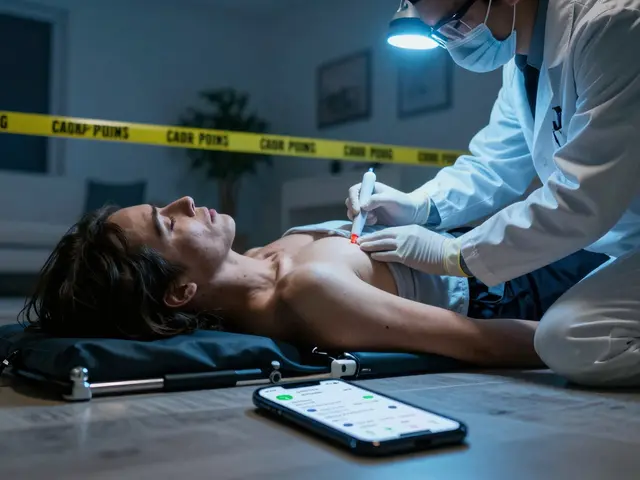
Fire scenes are chaotic. But when the flames die down and the body arrives on the autopsy table, the story isn't over-it's just getting complicated. You might see a victim who looks like they walked out of a movie set, skin intact, clothes barely singed, yet dead. Why? The answer usually lies in what you can't see with the naked eye: inhalation injury is damage to the airways and lungs caused by breathing in hot gases, smoke particles, or toxic chemicals. It kills more fire victims than the actual flame contact does.
If you're stepping into the morgue for a thermal death case, you need to know exactly where to look and what those marks mean. Misinterpreting a post-mortem artifact as an antemortem burn can flip a homicide investigation upside down. Or worse, miss a foul play entirely. Let’s break down how to read the body’s thermal history without guessing.
The Critical Distinction: Antemortem vs. Postmortem Damage
Before you pick up the scalpel, ask yourself one question: Did this person breathe while burning? This determines everything. A body thrown into a fire after death (postmortem) reacts differently than someone trapped inside (antemortem).
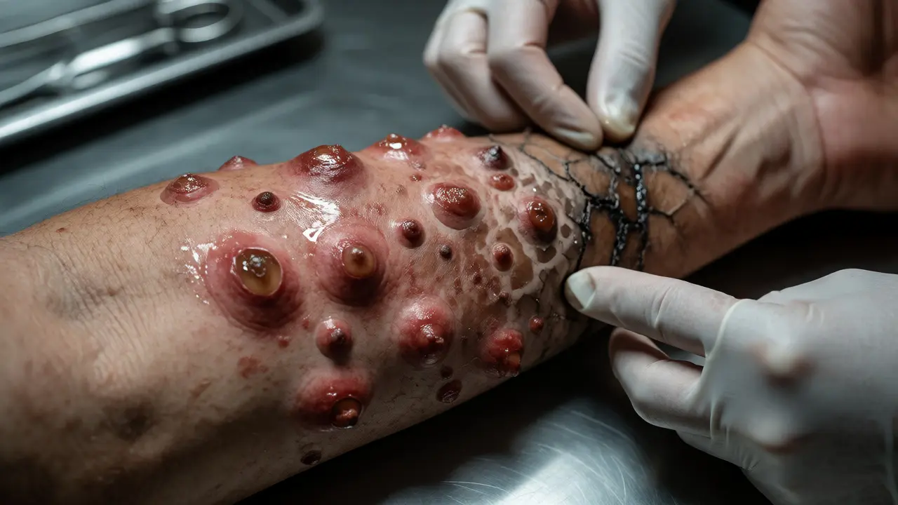
In living tissue, heat triggers an inflammatory response. You’ll see erythema (redness), edema (swelling), and blister formation filled with fluid that contains proteins and electrolytes. These blisters are gold mines for toxicology. If the fluid shows high levels of carbon monoxide or specific accelerants, you know the person was alive during exposure.
Conversely, postmortem burns lack these vital signs. The skin might char or split due to heat contraction, but there’s no swelling around the edges. No red line separating burned from unburned skin. If you find a "blister" on a corpse exposed to fire for hours, check the contents. If it’s mostly water or air bubbles, it’s likely heat-induced separation of skin layers, not a biological reaction. Always correlate histology with gross findings.
Decoding Burn Depth and Patterns
Forensic pathologists don’t classify burns just by depth; we look at pattern. The location tells you about position and movement. Did the victim try to escape? Were they sleeping?
Burn classification typically follows medical standards: first-degree affects only the epidermis, second-degree reaches the dermis, third-degree destroys all skin layers, and fourth-degree extends into muscle and bone. However, in autopsies, visual estimation is tricky because charring obscures layers.
Look for the "cleavage plane." In severe burns, the subcutaneous fat liquefies and separates from underlying structures. This creates gaps that can mimic trauma. Don’t mistake heat-induced separation for blunt force injury. Check the surrounding tissue for hemorrhage. True trauma bleeds; heat splits.
Also, watch for "candle wax" appearances. When synthetic fabrics melt onto skin, they create distinct, shiny patches. These patterns often map directly to clothing types. If you see a rectangular patch of melted polyester on the thigh, you’re looking at a shirt pocket or label. Documenting these helps reconstruct the victim’s attire, which matters if witnesses claim different clothes were worn.
The Silent Killer: Inhalation Injury Mechanics
Most people think fire kills by burning the outside. Wrong. Carbon monoxide poisoning and cyanide toxicity kill quickly, often before significant external burns appear. This is why a victim found far from the main fire source might still have fatal internal damage.
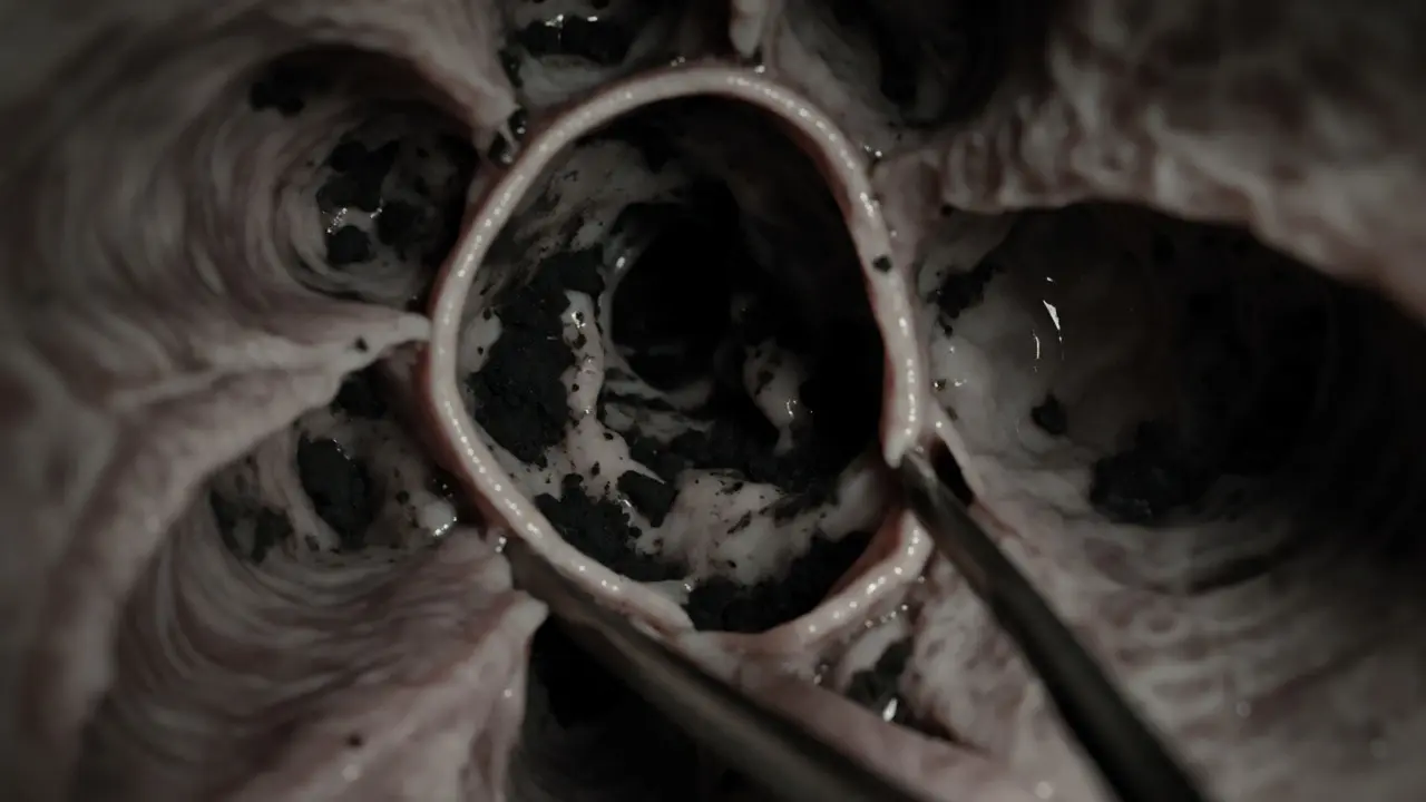
Smoke inhalation involves three mechanisms: thermal damage to the upper airway, chemical irritation to the lower airway, and systemic toxicity from gas absorption. The upper airway (nose, throat) acts as a heat exchanger. Unless the steam was superheated or the exposure prolonged, deep lung tissue rarely gets thermally burned. Instead, it suffers from soot deposition and chemical inflammation.
During autopsy, open the trachea and bronchi. Look for soot. Is it present above the vocal cords? Below them? Soot in the stomach confirms the victim swallowed smoke-laden mucus, proving survival past the initial exposure. If the stomach is empty of soot but the lungs are full, consider rapid incapacitation.
Cyanide adds another layer. Hydrogen cyanide forms when nitrogen-containing materials like wool, silk, or polyurethane foam burn. It blocks cellular oxygen use. Victims may have cherry-red skin (from CO) or normal-looking skin (cyanide). Test blood for carboxyhemoglobin (COHb) and cyanide levels. A COHb level above 50% is often lethal, but tolerance varies. Chronic smokers start with higher baselines, so interpret carefully.
Special Scenarios: Flashover and Confined Spaces
Flashover changes the game. It’s when a room suddenly ignites completely. Temperatures spike to over 1,000°F in seconds. Bodies in flashover scenarios often show uniform charring. There’s little variation in burn pattern because the heat hit everywhere at once.
In confined spaces, oxygen depletion plays a huge role. Fire consumes oxygen rapidly. Victims may suffocate before burning severely. You might find minimal external damage but massive pulmonary edema and congestion internally. This mismatch-low external burns, high internal distress-is a classic sign of oxygen-poor fire environments.
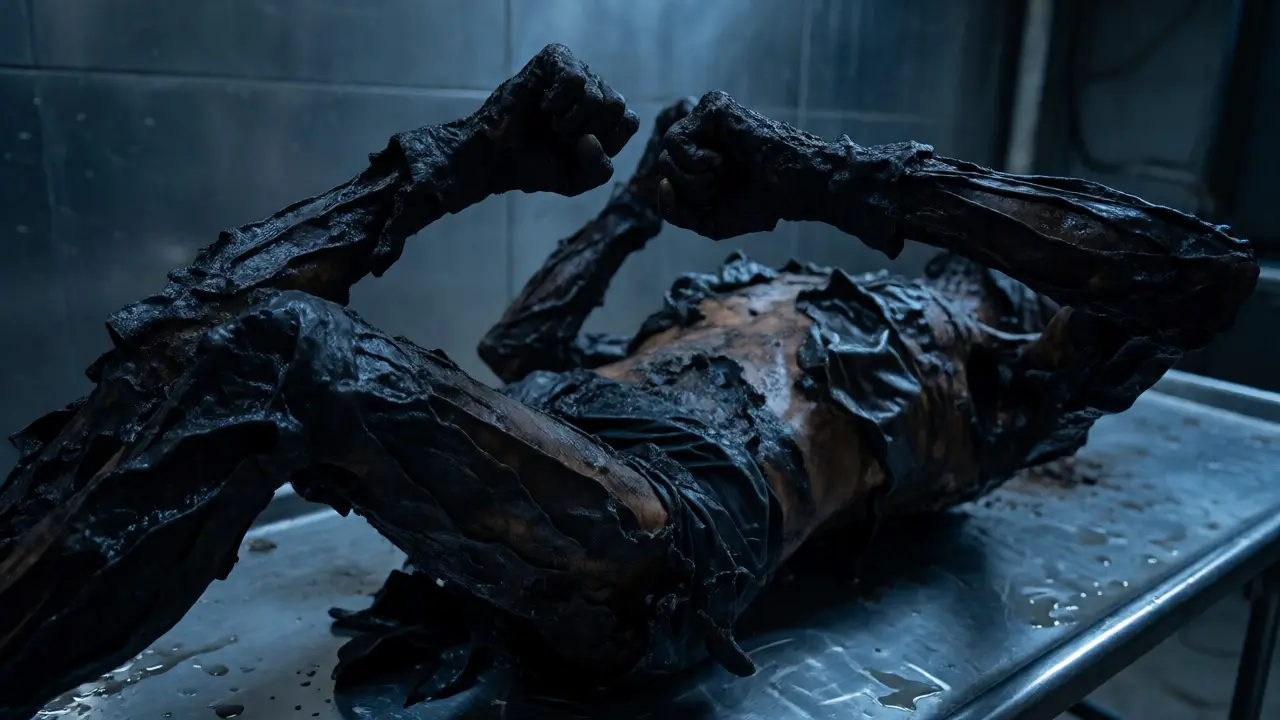
Consider the "pugilistic stance." As muscles dehydrate and contract from heat, limbs pull inward. Arms bend at elbows, knees draw up. This looks like fighting posture, leading families to believe their loved one struggled. Explain this clearly. It’s rigor mortis accelerated by heat, not combat. Document joint angles precisely to rule out pre-existing conditions or restraint effects.
Toxicology and Laboratory Analysis
Gross examination gives clues, but lab work provides proof. You cannot skip toxicology in any fire-related death.
| Marker | Source | Significance | Collection Note |
|---|---|---|---|
| Carboxyhemoglobin (COHb) | Carbon Monoxide | Indicates smoke inhalation; >50% often fatal | Draw peripheral blood to avoid contamination |
| Cyanide | Burning plastics/wool | Cellular hypoxia; difficult to detect post-mortem | Collect within 24-48 hours; unstable |
| Accelerants | Gasoline, kerosene, etc. | Suggests arson or accidental spill | Use inert containers; avoid plastic bags |
| Ethanol/Drugs | Prior consumption | Impairs escape ability | Vitreous humor stable even in decomposed bodies |
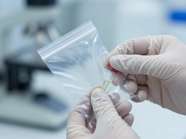
Note the collection notes. Peripheral blood is crucial for COHb. Central blood can be contaminated by diffusion from lungs. For cyanide, time is enemy. It breaks down fast. If you can’t run tests immediately, freeze samples properly. And never store accelerant evidence in plastic bags-volatiles leak through. Use glass jars lined with Teflon or clean metal cans.
Common Pitfalls and Artifacts
Even experienced pathologists get tripped up. Here are traps to avoid:
- Heat Fractures: Skull bones crack from heat expansion. These look like blunt force fractures. Check for hemorrhage. Heat fractures have dry, clean edges. Trauma has bleeding.
- Postmortem Blistering: Gas buildup under skin creates bubbles. They aren’t filled with serum. Pierce them. If they collapse flat, they’re artifacts.
- Color Changes: Pink discoloration can come from CO poisoning, cold exposure, or decomposition bacteria. Don’t assume cherry-red equals CO alone. Correlate with blood tests.
- Melted Evidence: Jewelry melts at different temperatures. Gold stays solid longer than aluminum. Finding melted zippers vs. intact buttons helps estimate temperature exposure duration.
Remember, the goal isn’t just to say "burned." It’s to determine cause and manner. Was it accidental? Suicidal? Homicidal? The distribution of burns, the presence of defensive wounds underneath soot, and toxicology results weave that narrative together.
Can a person survive a fire with no external burns?
Yes, absolutely. Many fire deaths result from smoke inhalation rather than direct flame contact. A victim trapped in a bedroom with closed doors might suffer fatal carbon monoxide poisoning or cyanide toxicity while remaining physically unburned. Autopsy must always include airway inspection and toxicology screening regardless of external appearance.
How do you distinguish antemortem from postmortem blisters?
Antemortem blisters contain protein-rich fluid and show inflammatory cells under microscopy. Postmortem blisters are caused by heat separating skin layers and contain mostly water or air bubbles without inflammatory markers. Histological analysis of the blister roof and fluid content is the definitive method for differentiation.
What is the pugilistic stance and why does it happen?
The pugilistic stance is a natural position adopted by burned bodies where arms flex at the elbows and knees draw toward the torso. It occurs because intense heat causes muscle dehydration and shrinkage, pulling joints into flexion. It is not a sign of struggle or defense but a physical reaction to thermal energy affecting soft tissues.
Why is peripheral blood preferred for COHb testing?
Peripheral blood is less susceptible to postmortem redistribution and contamination from lung gases. Central blood samples near the heart can falsely elevate COHb levels due to diffusion of carbon monoxide from the lungs into nearby vessels. Using femoral or brachial blood ensures a more accurate reflection of the victim’s status at the time of death.
Does the color of soot indicate the type of material burning?
Not reliably. While some materials produce distinctive smoke colors (e.g., black soot from incomplete combustion of hydrocarbons), soot composition varies based on oxygen availability and fuel mixture. Chemical analysis of soot deposits via mass spectrometry is required to identify specific sources, though visual inspection can hint at heavy particulate load versus lighter aerosol.