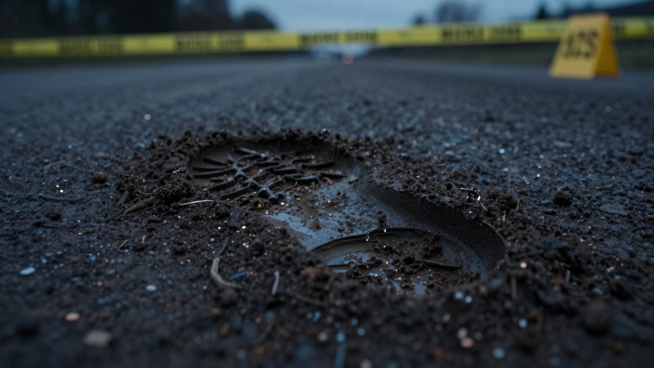
Imagine a suspect's boot prints at a crime scene. On the surface, it's just mud. But to a forensic geologist, that mud is a biological and chemical map. The real challenge isn't just finding the soil, but proving that the soil from a specific suspect's shoe matches a specific patch of ground at a crime scene. Because soil varies wildly over just a few meters, simple visual checks aren't enough. We need to look at the atomic and microscopic level to turn "dirt" into admissible evidence. That's where soil mineralogy comes into play, specifically using two heavy-hitting tools: X-ray diffraction (XRD) and scanning electron microscopy (SEM).
The Foundation of Soil Analysis
When we talk about soil in forensics, we aren't just talking about organic matter. We are looking at the inorganic crystalline structures-the minerals-that make up the earth. To get a definitive match in casework, analysts need to identify the specific mineral phases present. If two samples have the exact same rare mineral profile, the probability that they came from the same location skyrockets.
In a typical forensic workflow, the process starts with a basic microscopic exam. Before the high-tech machines are turned on, an analyst looks for "artifacts" like hair, seeds, or tiny pieces of plastic. Once those are cleared, the focus shifts to the mineralogical makeup. The goal is to find markers that are unique to a geographic area. While common minerals like Silica, a hard, crystalline compound of silicon and oxygen, Calcite, a carbonate mineral often found in limestone, and Hematite, an iron oxide mineral that gives soil a reddish hue are found in almost every sample, it's the rarer accessory minerals that provide the "smoking gun."
Unlocking Structure with XRD
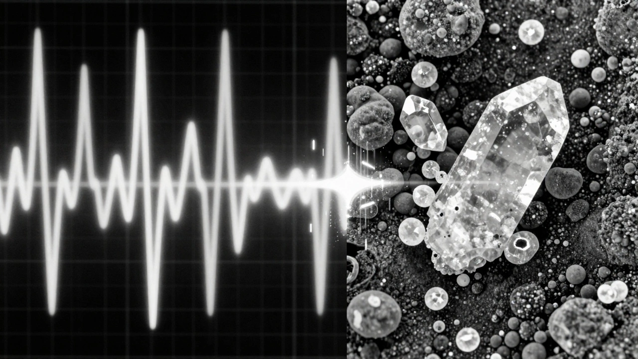
X-ray diffraction, or XRD is a non-destructive analytical technique used to identify the crystalline phases of a material by measuring the diffraction of X-ray beams, is the gold standard for bulk mineral identification. The beauty of XRD in forensic casework is that it's non-consumptive. In a legal setting, preserving evidence is everything. If a case goes to appeal five years later, the original sample must still exist. XRD allows us to get a chemical fingerprint without destroying the sample.
The process works in two main stages. First, the machine generates a diffraction pattern-a series of peaks. The analyst compares these unknown peaks against a massive database of known mineral patterns. However, soil is messy. It contains both crystalline (ordered) and amorphous (disordered) materials. To avoid mistakes, experts look at "d-spacing" values, which measure the distance between atomic planes. Even if two minerals look similar, their d-spacing is usually unique.
To keep things standardized, forensic labs follow ASTM E3294, the official standard guide for the forensic analysis of geological materials using powder X-ray diffraction. This ensures that whether the test is done in Oregon or New York, the method of dispersing minerals in aqueous solutions and segregating clay-sized materials is consistent and scientifically valid.
Adding Detail with SEM and EDS
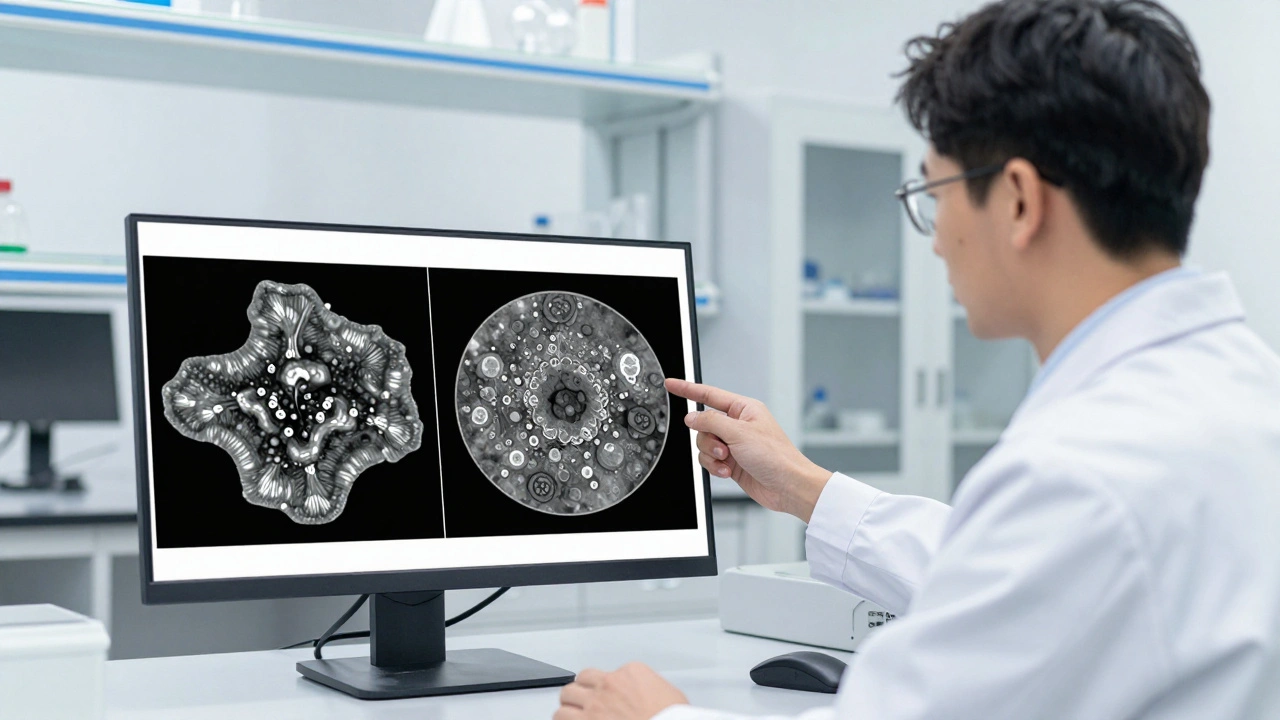
If XRD gives us the "what" (the list of minerals), SEM is Scanning Electron Microscopy, a technique that produces high-resolution images of a sample's surface by scanning it with a focused beam of electrons, gives us the "how" and "where." SEM doesn't just tell us that a mineral is present; it shows us the shape, size, and texture of the individual grains.
Most modern SEMs are equipped with EDS (Energy-Dispersive X-ray Spectroscopy). This allows the analyst to point the electron beam at a single, tiny grain and instantly determine its chemical composition. This is a game-changer for "modal mineralogy." While XRD analyzes the whole bulk of the soil, SEM can identify "lithotypes"-categories based on a combination of mineralogy, grain size, and how different minerals are associated with one another.
For example, if XRD tells you there is quartz and feldspar in both the crime scene sample and the suspect's shoe, that's a start. But if SEM shows that the crime scene quartz is rounded and weathered while the suspect's quartz is sharp and angular, you've just found a critical piece of evidence that differentiates the two samples.
| Feature | X-Ray Diffraction (XRD) | Scanning Electron Microscopy (SEM) |
|---|---|---|
| Primary Goal | Bulk mineral identification | Particle morphology & composition |
| Sample Impact | Non-destructive / Non-consumptive | Non-destructive (but requires coating) |
| Data Provided | Crystalline phases, d-spacing | Visual texture, grain size, EDS chemistry |
| Best For | Rapid screening & quantification | High-resolution discrimination |
| Perspective | Global (entire sample) | Local (individual grains) |
The Integrated Analytical Approach
In real-world casework, you don't choose one over the other; you use them as a team. The most effective strategy is a sequential workflow. First, XRD is used for a rapid bulk screen. This quickly tells the analyst if the samples are even remotely similar. If the XRD patterns are wildly different, the samples can be ruled out immediately without wasting more time.
If the XRD results are a match, the analyst moves to SEM. This is where they look for the nuanced differences in grain texture and mineral associations. By combining these two techniques with chemometrics-the use of mathematical models to analyze chemical data-forensic scientists can create a high-confidence match that can withstand the scrutiny of a courtroom.
The biggest pitfall to avoid is "sampling bias." Soil is not homogeneous. If you only take a tiny speck of dirt, you might miss the one rare mineral that defines the area. This is why systematic sub-sampling is required. Analysts must ensure that the particle size and modal abundance of the sample they test are representative of the larger soil mass.
From the Lab to the Courtroom
The transition from a laboratory finding to legal evidence requires a strict chain of custody and validated methods. Because XRD and SEM are widely accepted in the scientific community, they meet the criteria for reliability. When an expert witness testifies, they don't just say "the soil matches." They present the XRD diffraction peaks and the SEM imagery as physical proof of the mineralogical identity.
This level of detail prevents the common mistake of over-generalizing. Instead of saying "this is typical red clay," an expert can say "this sample contains a specific ratio of hematite and kaolinite with angular grain morphology consistent with the specific geological layer found at the crime scene." That level of specificity is what converts a lead into a conviction.
Is XRD truly non-destructive?
Yes. Unlike chemical digestion or some forms of mass spectrometry, XRD does not consume the sample. The X-rays interact with the crystalline lattice without altering the chemical structure, meaning the powder sample can be recovered and used for further testing or kept as evidence.
Why use SEM if XRD already identifies the minerals?
XRD identifies what is there, but SEM shows how it looks. Two soils can have the same mineral percentages but completely different grain shapes (e.g., rounded vs. angular) and textures. SEM allows analysts to see these physical differences, which are often the key to distinguishing two different locations with similar geology.
What is the role of EDS in soil analysis?
EDS (Energy-Dispersive X-ray Spectroscopy) is an attachment to the SEM that analyzes the X-rays emitted by a sample when hit by the electron beam. This allows the analyst to identify the elemental composition of a single specific particle, confirming its mineral identity in real-time.
How does ASTM E3294 help in forensic cases?
ASTM E3294 provides a standardized set of rules for analyzing geological materials. By following these guidelines, forensic labs ensure their methods are reproducible and can be defended in court, reducing the risk of the evidence being thrown out due to inconsistent lab procedures.
Can these techniques identify organic matter in soil?
XRD and SEM are primarily used for the inorganic (mineral) portion of soil. While SEM can see organic particles, it is not the primary tool for organic analysis. Forensic analysts typically use microscopy for vegetation and hairs, and other chemical methods for organic compounds.
Next Steps for Forensic Analysts
If you are integrating these techniques into a new lab workflow, start by establishing a local reference library of soils from your specific region. The effectiveness of XRD depends heavily on having accurate reference patterns to match against.
For those dealing with complex samples where minerals are heavily overlapped, consider implementing chemometric software. This helps in "deconvolving" the data, allowing you to separate overlapping peaks in an XRD pattern and get a cleaner reading of the mineral abundance. Finally, always ensure a rigorous sub-sampling protocol is in place to avoid the trap of sampling bias, which can lead to false exclusions in a criminal investigation.