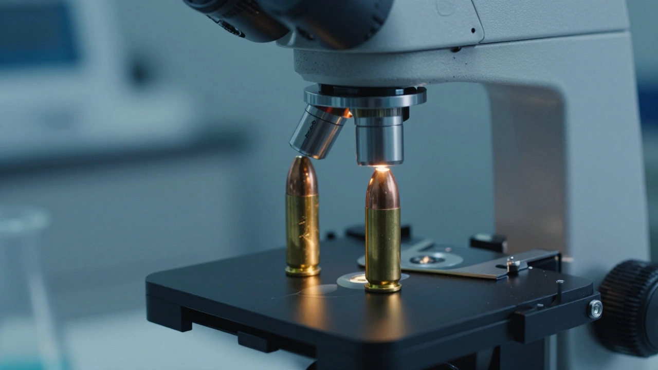
Imagine two bullets recovered from a crime scene. To the naked eye, they look like identical pieces of distorted lead. But under a specialized lens, they tell two completely different stories. The secret to unlocking these stories isn't just magnification-it's the ability to see two things at once. That is where Comparison Microscopy is an advanced optical technique that allows forensic specialists to examine two separate objects side-by-side through a single eyepiece. In the world of Bullet Analysis, this tool is the gold standard for proving whether a specific gun fired a specific shot.
The Science of the "Ballistic Fingerprint"
To understand why we need these microscopes, you first have to understand how a gun works. When a bullet travels through a barrel, it doesn't just slide through. The barrel has spiral grooves cut into it, known as rifling, which spin the bullet for stability. As the bullet pushes through, the harder metal of the barrel scrapes the softer metal of the bullet. This leaves behind microscopic scratches called Striations.
Here is the kicker: no two barrels are identical. Even if a company makes ten thousand guns of the same model using the same machinery, tiny imperfections in the steel and wear from use create a unique set of marks. These marks are effectively a fingerprint for the weapon. If a forensic examiner finds a bullet at a scene and then test-fires a suspected weapon into a water tank, they have two specimens. But remembering the exact pattern of a thousand tiny scratches while switching between two different microscopes is impossible. That is why the comparison microscope, with its optical bridge and split-view window, is non-negotiable.
Step-by-Step: The Analysis Process
Analyzing a bullet isn't as simple as just plopping two pieces of lead under a lens and hoping they match. It is a methodical process that ensures the evidence holds up in court.
- Preliminary Screening: Before the big microscope comes out, examiners use a Stereomicroscope. This gives a 3D view of the bullet to check for the quality of the marks and ensure the bullet isn't too deformed to be useful.
- Setting the Baseline: The evidence bullet is placed on the right stage. The examiner adjusts the lighting-usually using oblique illumination from the rear-to make the scratches pop. They scan the entire bearing surface at low magnification (10X or 20X) to find the most distinct area of characteristics.
- The Side-by-Side Match: Once a good area is locked in on the right, the test-fired bullet is placed on the left stage. The examiner then rotates the evidence bullet slowly. They are looking for the moment the "lands" (the raised parts of the barrel) and "grooves" (the recessed parts) align perfectly.
- Verification: If a match looks likely, the examiner switches to higher magnification. They rotate both bullets simultaneously to see if the fine striations continue to align across the entire surface. If the lines match up like a puzzle, you have a positive identification.
Class Characteristics vs. Individual Characteristics
Forensic experts divide their findings into two buckets: class and individual. Class characteristics tell you what kind of gun was used, while individual characteristics tell you which specific gun it was.
| Feature | Class Characteristics | Individual Characteristics |
|---|---|---|
| What it is | General design features from the manufacturer. | Random imperfections unique to one barrel. |
| Examples | Caliber, number of lands/grooves, twist direction. | Unique microscopic striations and nicks. |
| Purpose | Narrows down the make and model of the weapon. | Links a specific bullet to a specific firearm. |
| Consistency | Same for all guns of that model. | Different for every single gun. |
Beyond the Bullet: Cartridge Case Analysis
Comparison microscopy isn't just for bullets. The Cartridge Case (the brass shell) is often more valuable than the bullet itself. When a gun is fired, the firing pin slams the primer against the breech face of the chamber. This leaves a distinct impression. Additionally, the ejector and extractor mechanisms leave scratches on the rim of the case as it is kicked out of the weapon.
Examiners use the same split-screen approach to compare these marks. If a shell casing found at a crime scene matches the marks from a test-fired casing, it's a powerful piece of evidence, even if the bullet itself was destroyed or never recovered.
The Digital Shift: Virtual Comparison Microscopy
For decades, the only way to compare evidence was to have both the crime bullet and the suspect gun in the same room. This created a logistical nightmare. If the evidence was in New York and the gun was in Los Angeles, someone had to ship the evidence across the country, risking loss or damage.
Enter Virtual Comparison Microscopy (VCM). Instead of physical lenses, VCM uses high-resolution digital imaging. One lab can scan a cartridge case and send the digital file to another lab. The second lab can then overlay their own scan of a test-fire. This doesn't just save time; it adds a layer of digital archiving that traditional microscopy lacks. While the physical microscope is still the primary tool for final verification, VCM is rapidly becoming the first line of defense in multi-jurisdictional investigations.
Common Pitfalls and Challenges
It is easy to think this is a perfect science, but it has its hurdles. For instance, if a bullet hits a hard surface like concrete, it can "mushroom" or fragment. This distortion can wipe out the striations, making a match impossible. Similarly, if a barrel is cleaned with an abrasive brush or becomes heavily rusted, the internal characteristics can change over time, potentially altering the marks it leaves on a bullet.
Another challenge is the "twist direction." To determine this, an examiner holds the nose of the bullet pointing away from them. Depending on whether the impressions run to the left or right, they can identify the rifling twist. If the twist direction of the evidence bullet doesn't match the suspect gun, the examiner can immediately rule out that weapon without wasting hours on a detailed microscopic search.
Can two guns of the same model produce the same marks?
No. While they will share "class characteristics" (like the number of grooves), the microscopic "individual characteristics" are caused by random variations in the manufacturing process and subsequent wear and tear. This makes every barrel unique.
What happens if the bullet is too damaged to analyze?
If the bearing surface is too distorted, the examiner cannot establish a reliable match. In these cases, investigators shift their focus to the cartridge cases or other forensic evidence, as the bullet is considered "inconclusive."
How does a scanning electron microscope differ from a comparison microscope?
A comparison microscope is used for side-by-side visual matching of larger objects like bullets. A Scanning Electron Microscope (SEM) provides much higher magnification and is typically used to find tiny particles, such as gunpowder residue (GSR), rather than matching striation patterns.
Is virtual comparison as accurate as the physical method?
Digital imaging is incredibly precise and excellent for screening and collaboration. However, most forensic protocols still require a final physical verification using a traditional comparison microscope before the evidence is presented in court.
What are 'lands' and 'grooves' in ballistics?
Inside a rifled barrel, the 'grooves' are the channels cut into the metal, and the 'lands' are the raised portions between those channels. These two elements work together to grip the bullet and spin it.
Next Steps for Evidence Handling
If you are dealing with recovered ballistics, the priority is preservation. Never scrub a bullet to "clean it up" for the microscope, as you might accidentally scrub away the very striations the examiner needs. For those in the field, using a Stereomicroscope for an initial check is the best way to determine if a specimen is worth the time and resources of a full comparison microscopy session.